On this page
You hold a magnifier over a leaf and see a blur of green. That is usually technique, not the lens. Here is how to fix it, what each surface shows at 10x, how to sketch it, and where the lens stops being useful.
How do you use a hand lens on a leaf?
Most hand lenses magnify 5 to 20 times, and you should hold one about an inch in front of your eye, according to a hand lens guide from University of New Hampshire Extension. The guide recommends no more than 10x, because stronger lenses have a shorter focal distance and a smaller field of view.
Rest the hand holding the lens against your cheekbone.
Bring the leaf up to the lens, not your head down to the leaf. It usually focuses one to three inches from the lens.
Hold the leaf parallel to the lens and turn so light falls on it. A bent head shades the leaf.
Flip the leaf and repeat for the lower surface.
What can you see on the upper and lower surface of a leaf?
Use a broad leaf such as mango or hibiscus, and a grass blade. NCERT's Class 11 chapter Anatomy of Flowering Plants calls the dicot leaf dorsiventral, with reticulate (net) venation, and the monocot leaf isobilateral, with parallel venation. Look for:
Veins. On the broad leaf: the midrib, side veins and a net of finer veins around small green patches. On the grass blade: veins running side by side from base to tip. Also hold the leaf against a window.
Hairs (trichomes). NCERT calls epidermal hairs on the stem trichomes and says those of the shoot system are usually multicellular, branched or unbranched, soft or stiff, and sometimes secretory. They help cut water loss through transpiration. Note whether each surface has hairs, how crowded they are, and whether any end in a tiny bead.
Shine and edge. According to NCERT, the epidermis is often covered by a waxy cuticle. Check whether the margin is smooth, toothed or hairy.
What a lens usually will not show is stomata. According to NCERT, the lower (abaxial) epidermis of a dorsiventral leaf generally has more stomata than the upper (adaxial) one, which may have none. An isobilateral leaf has stomata on both surfaces. Checking this takes a microscope.
Why can't a hand lens show stomata?
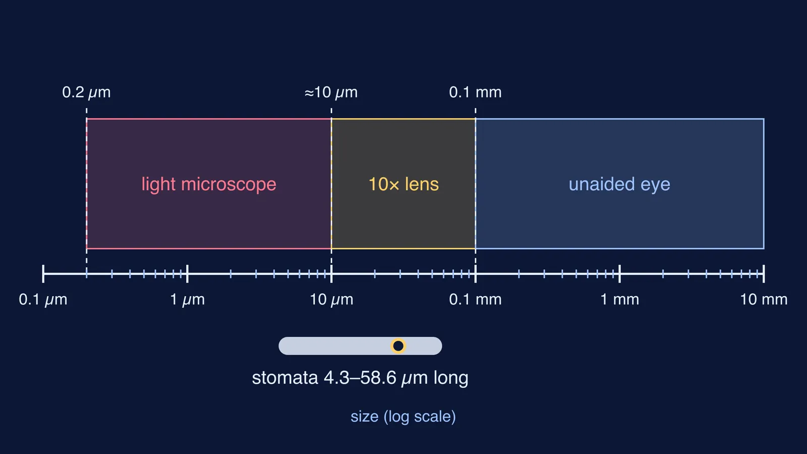
The unaided eye can resolve detail down to about 0.1 mm (100 µm), and a standard light microscope down to about 0.2 µm, according to a resolution primer from James Madison University's light microscopy facility. As a rough estimate, a 10x lens brings the eye's limit down to about 10 µm, which is 100 µm divided by 10.
A 2025 meta-analysis in New Phytologist pooled records from 89 species, with stomatal lengths from 4.3 to 58.6 µm. Stomata with kidney-shaped guard cells averaged 29.1 µm. An average stoma is only about three times the smallest detail a 10x lens separates, so at best you see a speck, not two guard cells and a pore.
How do you sketch leaf veins and hairs from a hand lens?
This plan follows the same habits as the biology record rules for drawing what you see.
Draw the outline large, about half a page, in sharp pencil with single, unbroken lines.
Draw the midrib as two lines tapering to the tip, and side veins as single lines at the angle you see. Count the side veins on one half and draw that number.
Don't draw every fine vein. Box one small patch and redraw it larger beside the leaf, labelled "detail, 10x hand lens".
In that detail box, draw hairs as short strokes showing direction and spacing. Write "branched" or "bead at tip" if you saw it.
Draw both surfaces side by side, labelled "upper (adaxial)" and "lower (abaxial)".
Add a scale line. The James Madison primer says a presented image needs a scale bar, because "Reporting the magnification is insufficient." Measure the leaf and mark what 1 cm is on your drawing.
Under each drawing, write one sentence comparing what you saw on the two surfaces.
If you mix up venation types, see types of phyllotaxy and venation with NCERT examples.
When do you need a microscope instead?
Choose the tool before you start:
Outline, margin, midrib and vein pattern: your eye, then a 10x lens.
Whether there are hairs, plus their spacing and shape on each surface: 10x lens.
The cells inside one hair: microscope.
Stomata, guard cells and stomatal counts: microscope and a leaf peel. See how to prepare a temporary mount of a leaf peel for stomata.
Palisade and spongy mesophyll: microscope and a transverse section.
If terms like dorsiventral, abaxial and trichome keep slipping, the Fermi Concept Library has a lesson for every biology concept, each with its place in the map, so you can check a term and see how it connects.
Take one leaf outside today, draw both surfaces and the detail box, then compare them with a peel under the microscope.