On this page
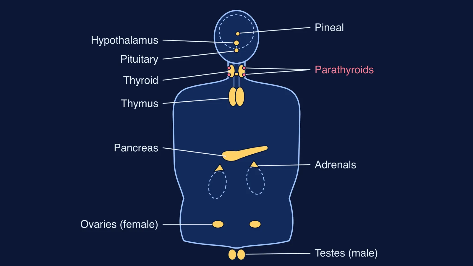
If a question asks you to label or draw the endocrine glands, you need one picture in your head: each gland next to a landmark you already know, such as the brain, the windpipe, the breastbone or the kidneys. The diagram above puts all of them on one outline, following Figure 19.1, "Location of endocrine glands", in NCERT Class 11 Biology, Chapter 19.
What are endocrine glands?
Endocrine glands lack ducts, so they are also called ductless glands, and their secretions are called hormones (NCERT Class 11 Biology, Chapter 19).
NCERT lists the pituitary, pineal, thyroid, adrenal, pancreas, parathyroid, thymus and gonads as the organised endocrine bodies, and describes the hypothalamus alongside them. The gut, liver, kidney and heart also make hormones, but they are not labelled on this diagram.
Where is each endocrine gland located in the human body?
Work from the top down. Each line gives the gland and its landmark, following the NCERT chapter unless another source is linked.
Hypothalamus: the basal part of the diencephalon, in the forebrain.
Pituitary: in a bony cavity called the sella turcica, attached to the hypothalamus by a stalk.
Pineal: on the dorsal (back) side of the forebrain.
Thyroid: two lobes, one on either side of the trachea, joined by a thin flap of connective tissue called the isthmus. The NCI SEER anatomy module adds that the lobes sit just below the larynx, or voice box.
Parathyroids: four glands on the back of the thyroid, one pair on each lobe.
Thymus: between the lungs, behind the sternum, on the ventral side of the aorta.
Adrenals: one pair, one above each kidney.
Pancreas: a long organ lying across the back wall of the abdomen, behind the stomach, from the duodenum to the spleen, according to the NCI SEER pancreas page.
Gonads: a pair of testes in the scrotal sac, outside the abdomen, in males; a pair of ovaries in the abdomen in females.
Which details do students mix up on the diagram?
A common slip is a gland drawn at the wrong landmark rather than a wrong name. These are the places to watch, all checked against the NCERT chapter.
Pituitary and pineal. The pituitary hangs below the hypothalamus on a stalk. The pineal is the other brain gland, on the dorsal side of the forebrain.
Parathyroids on the back. In a front view they are only dots at the edges of the thyroid lobes. NCERT shows them in a separate dorsal view, Figure 19.3 (b).
Thyroid or thymus. The thyroid is in the neck; the thymus is lower, in the chest behind the sternum.
Adrenals as caps. Draw each adrenal sitting above a kidney, not as a separate organ in the middle of the abdomen.
The pancreas is mostly not endocrine. NCERT says a normal human pancreas has about 1 to 2 million islets of Langerhans, making up only 1 to 2 per cent of pancreatic tissue. The rest is exocrine.
Testes outside the abdomen. They sit in the scrotal sac, below the outline of the abdomen; ovaries sit inside it.
Once the places are fixed, add what each gland secretes. Our guide to checking a gland-hormone-target table against NCERT helps with that, and parathyroid hormone vs thyrocalcitonin explains why the neck glands work as a pair on calcium.
How do you draw the endocrine system diagram from memory?
Use this five-minute drill on a blank sheet, a few times a week.
Outline: head, neck, chest, abdomen, pelvis. Sketch the brain, the windpipe and two kidneys as landmarks.
Brain trio: hypothalamus at the base of the brain, pituitary hanging below it on a stalk, pineal higher and towards the back.
Neck: a butterfly-shaped thyroid across the windpipe, just below the voice box, then four dots on its back for the parathyroids.
Chest: the thymus below the thyroid, behind the breastbone.
Abdomen: a cap on top of each kidney for the adrenals, then a long pancreas lying across behind the stomach.
Pelvis: two ovaries inside the abdomen, or two testes below it in the scrotal sac.
Label all nine, then compare with NCERT Figure 19.1. Circle each miss in a different colour.
Worked example: say you draw the thymus in the neck and the adrenals beside the pancreas. Write the landmark next to each miss (thymus: behind the sternum, between the lungs; adrenal: above the kidney) and redraw only those two the next day. When both land correctly twice in a row, stop drilling them.
If a miss keeps returning because the landmark itself is fuzzy (where is the diencephalon, or the duodenum?), fix the concept first. The Fermi Concept Library gives every biology concept its own lesson and its place in the map, and Fermi's AI tutor can draw the figure with you and check your understanding as it goes.
For more on this way of revising, see how to revise using textbook diagrams with the cover-and-check method, then try the drill once tonight.