On this page
Every living cell is enclosed by an outer boundary, but whether that boundary is a flexible gatekeeper or an unyielding wall depends on the organism. For students preparing for NEET-UG and board exams, confusing the cell wall with the cell membrane is a frequent source of lost marks. While both envelope the cell, their architecture, thickness, and permeability are fundamentally different.
What is the primary difference between a cell wall and a cell membrane?
The cell membrane is universal: every living organism, from single-celled amoebae to human nerve cells, relies on a plasma membrane to maintain homeostasis. By contrast, the cell wall is an extracellular layer found only in plants, fungi, bacteria, and most algae. Animal cells never form a cell wall, and specialised prokaryotes like Mycoplasma lack a cell wall entirely.
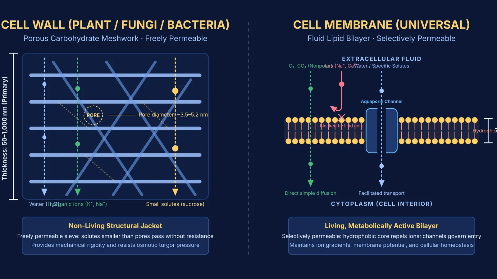
Their dimensions and vitality differ just as sharply. As documented by Daniel J. Cosgrove (2005), a growing primary plant cell wall measures between 50 and 1,000 nanometres in thickness, while secondary walls can exceed 2,000 nanometres. In contrast, the underlying plasma membrane described by S. J. Singer and Garth L. Nicolson (1972) is a microscopic lipid bilayer spanning only 7.5 to 10 nanometres. The cell membrane is living, fluid, and metabolically active; the cell wall is a rigid, non-living structural jacket secreted by the protoplast.
What are cell walls and cell membranes made of?
The chemical building blocks reflect their distinct roles. The cell membrane is organized as a fluid mosaic of lipids and proteins. In human erythrocytes, classical analysis cited by Singer and Nicolson (1972) reveals that the membrane consists of roughly 52 percent protein, 40 percent lipid, and 8 percent carbohydrates. Phospholipids form a bilayer with hydrophilic phosphate heads on the surfaces and hydrophobic fatty acid tails tucked inside.
Cell walls, by contrast, are carbohydrate nanocomposites:
In plants, walls consist of cellulose microfibrils embedded in hemicellulose and pectin, glued to neighbouring cells by the middle lamella (rich in calcium and magnesium pectate).
In fungi, the structural polymer is chitin, cross-linked with beta-glucans.
In bacteria, the wall is formed of peptidoglycan (murein), where glycan chains are cross-linked by short peptides.
For a kingdom-by-kingdom comparison, see our guide on what cell walls are made of in plants, fungi, and bacteria.
How do permeability and solute transport differ?
Permeability is where the functional contrast is starkest:
The cell wall is freely permeable. In living plant cells, Nicholas C. Carpita and colleagues (1979) measured the limiting pore diameter of primary walls at 3.5 to 5.2 nanometres. Because these pores are much larger than individual water molecules or inorganic ions, water, mineral salts, and small nutrients diffuse through without resistance.
The cell membrane is selectively permeable. Its hydrophobic lipid interior blocks charged ions and polar molecules. Small nonpolar gases diffuse across, but water moves rapidly through specialised channels called aquaporins, discovered by Gregory M. Preston and colleagues (1992). Ions and sugars require specific transport proteins and ATP.
How do cells respond to osmotic pressure with and without a wall?
The mechanical strength of the cell wall governs how cells react to osmotic water flux:
In a hypotonic medium (such as pure water), water enters down its potential gradient. Animal cells swell until the thin membrane bursts, causing osmotic lysis. Plant cells absorb water until the swelling protoplast pushes against the rigid wall, generating turgor pressure. The wall exerts an equal opposing wall pressure, stopping further water entry and preventing rupture.
In a hypertonic medium, water leaves the cell. Animal cells shrink and crenate. In plant cells, the rigid wall retains its shape while the plasma membrane pulls away from the wall—a process called plasmolysis.
The wall also dictates how dividing cells separate, which you can explore in our review of cytokinesis in plant and animal cells.
How can you test your understanding before exam day?
Use this four-step diagnostic checklist when tackling boundary questions in biology exams:
Step 1: Check the kingdom. Animal tissues and wall-less bacteria like *Mycoplasma* never have a cell wall.
Step 2: Check the thickness. A dimension of 7.5 to 10 nanometres (as established by Singer and Nicolson (1972)) indicates a lipid bilayer; a thickness above 50 nanometres (as detailed by Cosgrove (2005)) points to an extracellular wall.
Step 3: Check permeability. Unrestricted diffusion of small solutes flags a cell wall, while active transport or channel-mediated flow flags a cell membrane.
Step 4: Check osmotic response. If immersion in pure water generates turgor without lysis, the cell has a protective wall.
When solving numerical problems on water potential and turgor pressure, the Fermi Problem Library provides worked problems with a step-by-step solver that explains the underlying reasoning rather than just printing an answer. If you are reviewing cellular architecture from first principles, the Fermi Concept Library offers lessons that map every concept across biology and the physical sciences.