On this page
Cut DNA with a restriction enzyme and you get pieces you cannot see. Gel electrophoresis shows how many there are and roughly how long each is. The habit that makes it stick: predict the band pattern first, then check it against a ladder.
Why do smaller DNA fragments travel farther in a gel?
DNA fragments are negatively charged, so in an electric field they are pulled towards the anode, the positive electrode, as the NCERT Class 12 Biology chapter on biotechnology explains. The charge comes from DNA's phosphate backbone, according to a methods paper by Pei Yun Lee and colleagues in the Journal of Visualized Experiments.
The agarose gel acts as a sieve, and NCERT puts it plainly: "the smaller the fragment size, the farther it moves."
The key rule for reading sizes: "the distance traveled is inversely proportional to the log of its molecular weight", in the words of Lee et al., 2012. In practice, distance drops roughly in a straight line as log(size) rises, so every doubling of fragment length takes about the same fixed distance off a band's run; it does not halve it.
To see the bands, NCERT notes, the gel is stained with ethidium bromide and viewed under UV light, where DNA shows up as bright orange bands.
Predict first: which fragment travels farthest?
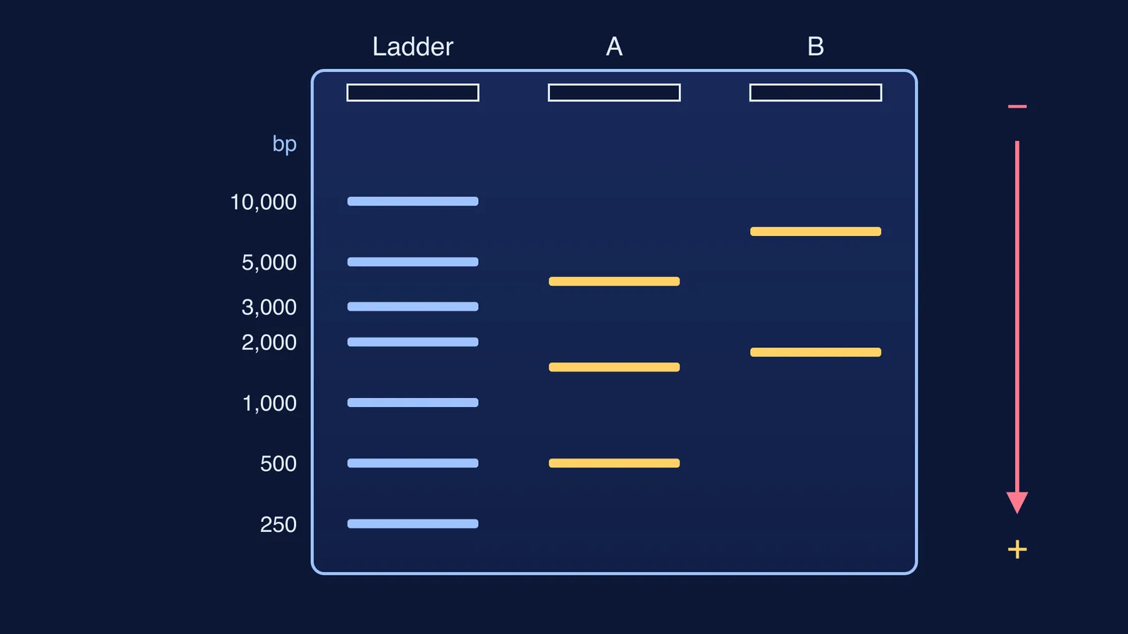
A linear 6,000 bp piece of DNA is cut into fragments of 4,000, 1,500 and 500 bp, loaded in lane A beside a ladder. Write your answers down.
How many bands will lane A show?
Which band sits closest to the wells?
Which band runs farthest?
Is the 1,500 bp band nearer the 500 bp band, or about halfway between the other two?
Answers: three bands; 4,000 bp nearest the wells; 500 bp farthest. Question 4 is the trap. In base pairs, 1,500 is much closer to 500 than to 4,000, but on a log scale it lands close to halfway, slightly nearer 4,000. The diagram at the top of this post is this gel drawn to scale.
Fragments of equal length share one band, so you can see fewer bands than fragments. Our guide to how many fragments a restriction enzyme makes walks through the counting step.
How do you read a fragment's size from a DNA ladder?
A ladder is a mix of DNA fragments of known sizes run in its own lane, and Lee and colleagues say a size marker "should always be loaded along with experimental samples" (JoVE methods paper). Thermo Fisher's GeneRuler 1 kb ladder, for example, has 14 fragments covering 250 to 10,000 bp (product page).
Worked example (practice distances from the wells, following the log rule):
10,000 bp: 10.0 mm
5,000 bp: 16.0 mm
3,000 bp: 20.5 mm
2,000 bp: 24.0 mm
1,000 bp: 30.0 mm
500 bp: 36.0 mm
250 bp: 42.0 mm
An unknown band in lane B is at 25.0 mm. To size it:
Find the ladder bands on either side: 2,000 bp at 24.0 mm and 1,000 bp at 30.0 mm.
Work out how far along that gap the unknown sits: (25.0 - 24.0) / (30.0 - 24.0) = 1/6.
Move the same fraction along the log scale. log10(2,000) = 3.301 and log10(1,000) = 3.000, so log10(size) = 3.301 - (1/6)(0.301) = 3.251.
Undo the log: 10^3.251 is about 1,780 bp.
If you interpolate in plain base pairs instead, you get 2,000 - (1/6)(1,000), about 1,830 bp. The error grows near the wells. Lane B's second band, at 13.0 mm, sits halfway between the 10,000 and 5,000 bp bands: it reads about 7,070 bp on the log scale but 7,500 bp on a straight one.
For a whole gel, plot log10(size) against distance for every ladder band, draw the best straight line, and read each unknown off it.
What changes how far DNA fragments travel?
Agarose concentration. Most gels are 0.5% to 2% agarose, with the concentration chosen to suit the fragment sizes being separated; the higher the concentration, the smaller the pores (Lee et al., 2012).
Voltage and run time. The same protocol sets the power supply to 1 to 5 V/cm between the electrodes. A longer or stronger run moves every band farther, ladder included, so compare bands only within one gel.
A predict-then-check routine for gel questions
List the fragment sizes the digest produces.
Merge equal sizes into one band.
Rank them: largest nearest the wells, smallest farthest.
Sketch the lane before you look at the gel or the answer key.
If your sketch is wrong, name the step that failed: the cutting, the merging or the ranking.
The Fermi Simulation Lab has interactive simulations for ideas that are easier to see than to read, and the same predict-then-test habit works there. If digest questions keep catching you out, Fermi's AI tutor can work out what you already know and build a plan for the session.
Next, read how gel electrophoresis separates and shows DNA fragments and how EcoRI cuts DNA and leaves sticky ends.