

On this page
If you are learning about biotechnology for your school board exams, restriction enzymes are introduced as the "molecular scissors" of the cell. They do not just chop DNA at random; they scan the double helix to find a specific sequence of letters and make a precise, predictable cut. This discovery opened the door to genetic engineering, allowing scientists to build recombinant DNA and mass-produce medicines like human insulin.
The most famous of these enzymes is EcoRI. But how exactly does it work, and what do its "sticky ends" look like when you draw them out?
What is a restriction enzyme?
A restriction enzyme, or restriction endonuclease, is a protein that cuts DNA at a specific recognition site. Bacteria originally evolved these enzymes as a defence mechanism against viruses (bacteriophages). When a virus injects its DNA into the bacterium, the restriction enzyme chops the foreign viral DNA into harmless pieces before it can take over the cell.
To protect its own DNA from being cut, the bacterium adds methyl groups to its own recognition sequences. This stops the enzyme from binding.
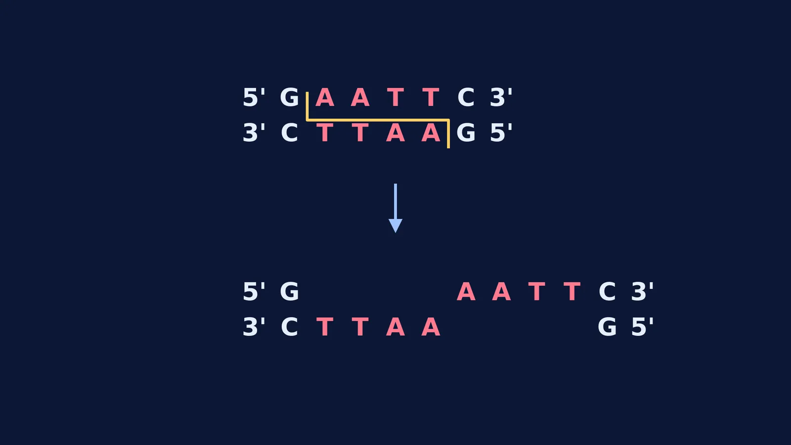
Each restriction enzyme recognises a different sequence. EcoRI was isolated from the bacterium *Escherichia coli* (strain RY13). It specifically looks for the six-letter sequence GAATTC (Hedgpeth et al., 1972). When it finds it, it cuts both strands of the DNA double helix.
However, EcoRI does not cut straight down the middle. It makes a staggered cut between the guanine (G) and adenine (A) on both strands.
Why do restriction enzymes leave sticky ends?
Because the DNA double helix has two strands running in opposite directions (antiparallel), the GAATTC recognition sequence reads the same forwards on the 5' to 3' strand as it does on the complementary 3' to 5' strand. This is known as a palindromic sequence. In everyday language, a palindrome like "racecar" reads the same forwards and backwards. In DNA, it reads the same when you flip to the opposite strand and read in the same 5' to 3' direction.
When EcoRI makes its staggered cut between the G and the A on both strands, the two pieces of DNA pull apart. One piece is left with a single-stranded tail of AATT on the top, and the other piece has an identical tail of TTAA on the bottom.
These exposed single strands are called "sticky ends" because they are eager to form hydrogen bonds with a complementary sequence. If you cut a human target gene and a bacterial plasmid with the same EcoRI enzyme, they will both have the exact same AATT sticky ends.
When you mix the human gene and the plasmid together in a tube, their matching ends find each other and "stick" together via base pairing. Finally, another enzyme called DNA ligase is added to seal the breaks in the sugar-phosphate backbone, creating a continuous piece of recombinant DNA.
How do you predict the fragments when EcoRI cuts a plasmid?
When you face exam questions about restriction enzymes, you are asked to predict how many fragments will form and how long they will be in base pairs (bp). Here is a framework to solve these problems without confusion.
Count the restriction sites: A linear piece of DNA cut once will give two fragments. However, a circular plasmid cut once will just open up into a single linear fragment. A plasmid cut twice will give two fragments.
Trace the cuts: If a 5000 bp plasmid has an EcoRI site at position 1000 and another at position 3500, the enzyme will cut at both spots.
Subtract to find the lengths: The first fragment is the distance between the two sites: 3500 − 1000 = 2500 bp. The second fragment is the rest of the circle: 5000 − 2500 = 2500 bp.
Write out the overhangs: If the question asks you to draw the product, write out the GAATTC sequence, draw a line between G and A, and clearly show the 5' AATT 3' overhang.
Reading about sticky ends in a textbook can still leave you guessing, because manipulating DNA is a physical, 3-dimensional process. The Fermi Simulation Lab has interactive simulations for the ideas that are easier to see than to read. You can load a virtual plasmid, run an interactive restriction enzyme simulation, and watch the DNA split to form sticky ends. You can then try to paste in a target gene and check if the ends match up correctly.
What happens after the sticky ends are joined?
Once you have successfully cut and pasted a gene into a plasmid, you have created recombinant DNA. The next step is to introduce this new plasmid into a host bacterium, so that the cell can read the gene and manufacture the protein you want.
This process requires chemical or physical methods for getting DNA into host cells, such as heat shock or microinjection. When you can accurately picture the staggered cut of EcoRI and the matching of sticky ends, the entire biotechnology syllabus makes logical sense.


