On this page
Most students meet DNA as a flat ladder on a textbook page. That picture hides what exam questions keep testing: how the pairs fit inside a twisted helix, and why the strands point opposite ways. Here you'll turn the helix like a model in your hands, then predict partners and count hydrogen bonds on a real structure.
What does the DNA double helix look like in 3D?
NCERT's Class 12 chapter Molecular Basis of Inheritance lists the salient features. Each of the two chains has a sugar-phosphate backbone, and the bases project inward. The pitch of the helix is 3.4 nm, with roughly 10 base pairs in each turn, so neighbouring base pairs sit about 0.34 nm apart. The two chains coil in a right-handed fashion. Stacking of one base-pair plane over the next adds to the stability that hydrogen bonds give.
The same numbers appear in the original model. In their April 1953 Nature paper, James Watson and Francis Crick put "the bases on the inside of the helix and the phosphates on the outside", with a residue every 3.4 Å and a repeat after 10 residues, or 34 Å (3.4 nm).
When you rotate a model, check three views:
Side-on: two backbones wind round the outside, with base pairs as rungs. Count about 10 rungs per full turn.
Down the axis: the pairs stack like a pile of coins.
End over end: flip it 180° about a line across its middle. The backbones swap places. Remember this view for the last section.
Which base pairs with which, and how many hydrogen bonds?
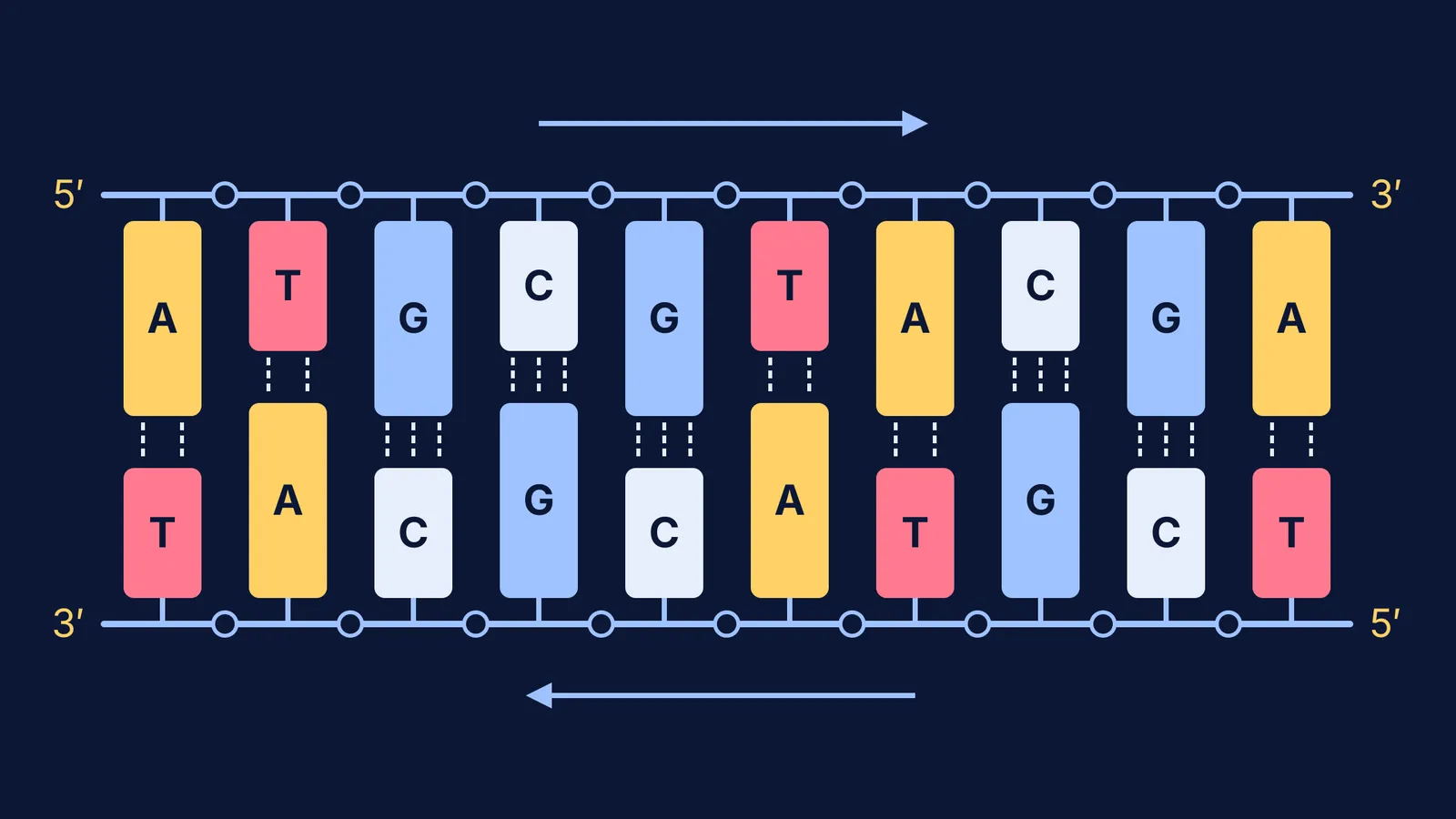
According to NCERT, adenine forms two hydrogen bonds with thymine, and guanine forms three hydrogen bonds with cytosine. Adenine and guanine are purines; thymine and cytosine are pyrimidines. So a purine always faces a pyrimidine, which NCERT says keeps the distance between the strands almost uniform. In the diagram at the top of this page, long and short blocks add up to the same width at every rung. The pairing also explains Chargaff's rule, as NCERT states it: in double-stranded DNA, A:T and G:C ratios both equal one.
The G–C count took a few years to settle. In 1956, Linus Pauling and Robert Corey90185-0) published on specific hydrogen bonding between purines and pyrimidines, and in a September 1957 letter to Crick, Pauling wrote that they had shown guanine and cytosine share three hydrogen bonds, not two.
Worked example: predict the partner strand and count the bonds
Take the top strand from the diagram: 5′-ATGCGTACGA-3′.
Write the partner under each base, using A↔T and G↔C: 3′-TACGCATGCT-5′.
Turn it round so it reads 5′→3′, as answers usually expect: 5′-TCGTACGCAT-3′.
Count the pairs. A–T pairs: 5. G–C pairs: 5.
Hydrogen bonds: 5 × 2 + 5 × 3 = 25.
Now try it on real data. Entry 1BNA in the RCSB Protein Data Bank is a B-DNA dodecamer from Drew, Dickerson and colleagues, released in 1981, with each chain reading CGCGAATTCGCG. Its page has a 3D viewer you can rotate in the browser. Predict the partner first: it is CGCGAATTCGCG again, read 5′→3′, because the sequence is its own reverse complement. With 8 G–C and 4 A–T pairs, the two-and-three rule gives 8 × 3 + 4 × 2 = 32 hydrogen bonds.
Common slips to check for:
Writing the partner 5′→3′ without reversing it.
Putting uracil in DNA; NCERT notes it replaces thymine in RNA.
Counting hydrogen bonds per base instead of per pair.
Misusing Chargaff: if adenine is 20% of the bases, thymine is 20%, G and C share 60%, so cytosine is 30%.
If base pairing still feels shaky, the Fermi Concept Library has a lesson for every concept in biology and chemistry, each with its place in the map.
Why do the strands run antiparallel?
Each chain has a direction. NCERT describes a free phosphate on the 5′ carbon of the sugar at one end and a free OH on the 3′ carbon at the other. In the double helix, one chain runs 5′→3′ and its partner 3′→5′.
Watson and Crick gave the geometric reason: both chains follow right-handed helices, "but owing to the dyad the sequences of the atoms in the two chains run in opposite directions". A dyad is a two-fold axis: the line you flipped the model around. Turning 180° about it moves chain one into chain two's place and swaps its 5′ and 3′ ends, so the chains must run opposite ways. That direction matters again when DNA is copied, as our comparison of DNA replication and transcription shows point by point.
To go further, see how 3D molecule viewers help in chemistry, and revise the rest of the NEET chapter with our guide to the molecular basis of inheritance. For other ideas that are easier to see than to read, the Fermi Simulation Lab has interactive simulations you can try.