On this page
In short
Roger Tsien and his laboratory expanded the single green colour of the jellyfish fluorescent protein (GFP) into a full palette by mutating its structure. By engineering proteins like the red mCherry and far-red mPlum, they allowed scientists to tag and track multiple molecules at once inside living cells without the colours overlapping.
The inside of a living cell is a crowded, colourless place. When Osamu Shimomura isolated the green fluorescent protein (GFP) from a jellyfish, and Martin Chalfie demonstrated that it could be attached to other proteins, biology gained a way to see where molecules go. But GFP only glowed green. If you wanted to watch two different proteins interact, or see a virus enter a nucleus, a single colour was not enough.
What did Roger Tsien discover?
Roger Tsien shared the 2008 Nobel Prize in Chemistry for expanding the fluorescent palette. His laboratory at the University of California, San Diego, altered the genetic sequence of GFP to change the shape of its chromophore—the part of the protein that absorbs and emits light. By tweaking the amino acids around the chromophore, Tsien created blue, cyan, and yellow fluorescent variants, giving researchers their first multi-colour tools.
What are the mFruit fluorescent proteins?
Tsien's lab soon looked beyond GFP for longer wavelengths, finding a starting point in a red fluorescent protein called DsRed from a *Discosoma* coral. However, DsRed naturally clumped together in groups of four, forming a tetramer that ruined the function of any protein it was attached to. In 2002, Tsien's team used directed evolution to break the tetramer apart, creating mRFP1, the first true monomeric red fluorescent protein, which matured over 10 times faster than the original DsRed.
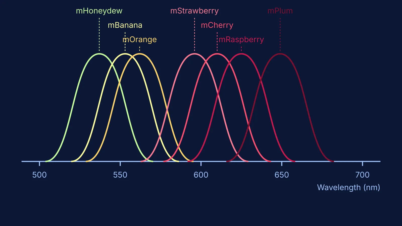
Over the next two years, the lab subjected mRFP1 to further mutations, yielding a rainbow of new proteins named after fruits that matched their colours. The 2004 "mFruit" palette included mHoneydew, mBanana, mOrange, mStrawberry, and mCherry. Another technique, borrowing the hypermutation process of immune cells, produced mRaspberry and mPlum.
Why do we need different colours of fluorescent proteins?
Distinct colours allow for simultaneous tracking. A researcher can tag the nucleus blue, the mitochondria green, and a structural protein red, then watch how they behave under a microscope without the signals overlapping. This is the same principle that allows Stefan Hell and the microscope that broke the diffraction limit to map structures at the nanoscale.
Longer wavelengths also penetrate tissue better. While blue and green light scatter easily and can damage living cells, the far-red light emitted by mPlum (at 649 nm) passes through biological tissue with far less interference, letting scientists look deeper into an organism.
How do we track the cell's machinery?
Because these tags are genetically encoded, you do not need to inject a dye. You simply splice the gene for mCherry next to the gene for your protein of interest, and the cell builds the glowing flag itself. This technique has mapped everything from George Palade and the ribosomes in the cell's protein factory to the intricate wiring of the brain.
If you are learning how changes in a DNA sequence translate to the physical shape of a protein, the Fermi Concept Library contains every concept in physics, chemistry, biology, mathematics and computing, each with its own lesson and its place in the map.
Tsien's engineering of the mFruit series turned a natural curiosity into one of the most powerful toolkits in modern biology, proving that sometimes, the best way to understand life is to colour it in.