

On this page
In the seventeenth century, botanists classified plants almost exclusively by their exterior leaves, petals, and herbal uses. While Robert Hooke's 1665 observation of cork cells had revealed empty cavities in dead bark, no one had mapped how living vegetation was constructed. English physician and botanist Nehemiah Grew changed that. Holding degrees from Cambridge and Leiden University, Grew approached plants with a medical eye, suspecting that they possessed internal organs just as animals do (Wikipedia). By spending more than a decade dissecting stems, roots, seeds, and blossoms under the compound microscope, he fundamentally shifted how scientists understood plant life.
In 1682, Grew published his *magnum opus*, *The Anatomy of Plants*, illustrated with 83 detailed copperplate engravings (Notes and Records of the Royal Society). The volume consolidated four earlier treatises presented to the Royal Society, establishing plant morphology as an empirical science (Notes and Records of the Royal Society). Alongside Italian physician Marcello Malpighi, who was conducting similar research independently in Bologna, Grew became recognized as the co-founder of plant anatomy (Encyclopaedia Britannica).
What did Nehemiah Grew discover under the microscope?
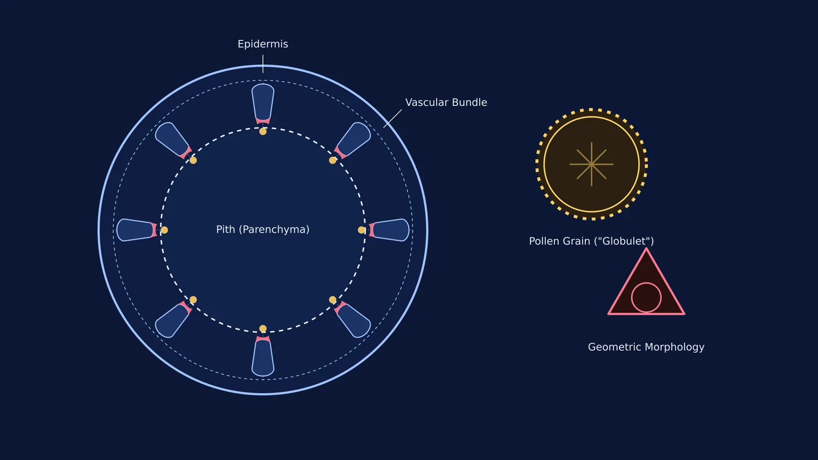
Before Grew, naturalists viewed stems and roots as uniform, solid cylinders. Slicing thin cross-sections with a razor, Grew placed them under his lenses and observed that plant matter resolved into a precise architecture of tubes, pores, and concentric zones. He identified key differences between stem and root morphology. He noted that roots form a dense central core of conducting vessels, which acts like a strong cable to anchor the plant in the soil, while stems arrange their vascular bundles toward the outside to provide structural rigidity (Wikipedia).
Grew also identified medullary rays radiating from the central pith, comparing the plant's structure to "fine bone-lace" where vertical fibers and horizontal tissues weave together. He coined enduring botanical terms, introducing "radicle" for the embryonic root and "plume" (now plumule) for the embryonic shoot (Encyclopaedia Britannica).
How did Grew describe plant tissues and parenchyma?
Grew's defining histological contribution was naming the fundamental ground tissue of plants. Borrowing the Greek term *parenchyma* from his training in animal anatomy, he used it to describe the soft, unspecialized tissue forming the pith, cortex, and pulp of fruits (Encyclopaedia Britannica). Under his lenses, this parenchyma appeared as countless tiny chambers or "bladders." He struggled to find the right words to convey this microscopic world, eventually writing that it resembled "the Froth of Beer or Eggs" or a "fine-wrought Sponge" (Notes and Records of the Royal Society).
How did Grew identify pollen and plant sex organs?
In Book IV of *The Anatomy of Plants*, Grew turned his microscope to flowers, leaves, and fruits. He provided the first microscopic account of pollen grains, calling them "globulets" that were scattered upon the attire of the flower (Encyclopaedia Britannica). While pollen seems like uniform yellow dust to the naked eye, Grew found that grains possess distinctive geometric forms—some smooth, others spiky or ridged—that remain remarkably constant within a single species. This observation established the foundation for modern palynology and our understanding of microsporogenesis and pollen grain structure.
Grew also published the first anatomical case for plant sexuality. Prompted by Oxford physician Sir Thomas Millington, Grew showed that the stamen, or "attire," produces pollen to fertilize the pistil (Encyclopaedia Britannica). By identifying stamens as male reproductive organs, Grew laid the observational groundwork for Rudolf Camerarius's experimental proof of plant pollination in 1694.
How can students read a plant tissue cross-section today?
Reading transverse sections under a microscope remains a core practical skill in Class 11 and NEET botany. Just as Grew did in 1682, you can identify any unknown plant section using a 4-step framework:
Check the outer boundary: A thick waxy cuticle with multicellular hairs indicates a stem; a thin epiblema with unicellular root hairs indicates a root.
Inspect the cortex: Stems feature a distinct hypodermis (collenchyma in dicots, sclerenchyma in monocots), while roots display a broad parenchymatous cortex ending at an endodermis with Casparian strips.
Map the vascular bundles: Dicot stems show bundles in a neat ring; monocot stems show scattered bundles; roots show alternating radial bundles.
Determine xylem maturation: Inward-pointing protoxylem (endarch) indicates a stem; outward-pointing protoxylem (exarch) indicates a root.
For a complete breakdown of these diagnostic checks, see our guide to identifying any flowering plant transverse section. When students transition from textbook diagrams to actual microscope slides, the Fermi Concept Library places every tissue into an integrated biological map. If you are practicing slide identification or floral formulas, Fermi's AI tutor works through problems alongside you, checking your reasoning at each layer rather than simply printing an answer.


