On this page
Is a brain area's job set by its genes, or by the signals it receives? Mriganka Sur tested this by sending signals from the eye into the part of the brain that normally handles sound, then checking whether that region learned to see.
Sur studied electrical engineering at IIT Kanpur and did his PhD at Vanderbilt University. He is now Newton Professor of Neuroscience at MIT and a Fellow of the Royal Society, according to his MIT faculty profile.
How did Mriganka Sur rewire a ferret's brain?
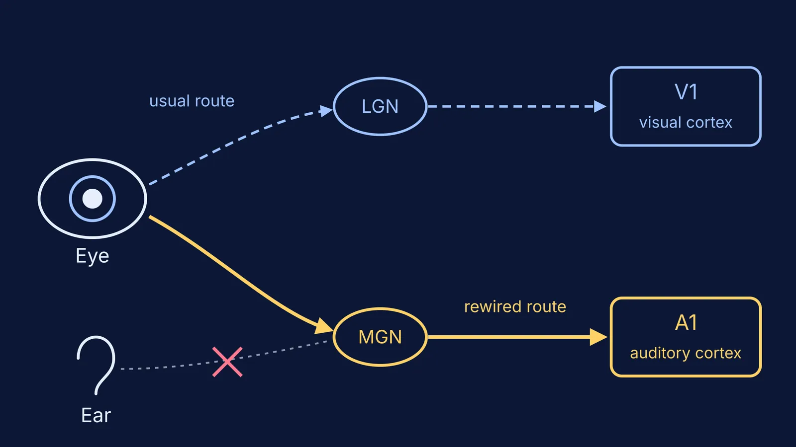
In a normal mammal, fibres from the retina carry visual signals to a relay station in the thalamus called the lateral geniculate nucleus (LGN). The LGN passes them on to the primary visual cortex (V1). Sound takes a parallel route: from the ear, through relay stations in the brainstem, to another thalamic relay, the medial geniculate nucleus (MGN), and then to the primary auditory cortex (A1).
Ferrets are born with these connections still forming, so Sur's team worked on newborn animals. In a 1988 paper in Science, Sur, Preston Garraghty and Anna Roe reported that they induced retinal cells to project into the MGN by reducing the usual targets of retinal axons in one hemisphere and creating alternative space for them in the auditory thalamus. Many MGN cells then responded to light, and some visual cells in the auditory cortex were tuned to the direction of motion or to tilted edges, much like cells in visual cortex.
Did the hearing cortex really learn to see?
Two papers in the 20 April 2000 issue of Nature answered this from two sides.
The first looked at wiring. Visual cortex is famous for orientation modules: groups of cells that prefer edges at the same angle, arranged into a map. Jitendra Sharma, Alessandra Angelucci and Sur found the same kind of modules in rewired auditory cortex. The neurons were tuned to orientation about as sharply as cells in V1, but the map was less orderly. In their data, the map's "pinwheel centres" were about one-quarter as dense in rewired A1 (1.1 per square millimetre) as in V1 (4.5 per square millimetre) (Sharma, Angelucci and Sur, Nature 2000).
The second looked at behaviour. Laurie von Melchner, Sarah Pallas and Sur tested rewired ferrets with light shown only in the part of the visual field "seen" by the rewired pathway. The animals responded as though they perceived the stimuli to be visual rather than auditory. They could also tell apart striped gratings of different spatial frequencies, though with lower acuity than the normal visual pathway.
The authors' conclusion: the perceptual modality of a region of cortex is instructed to a significant extent by its extrinsic inputs.
What does this tell us about brain plasticity?
Plasticity means the brain's wiring changes with activity and experience. Sur's work showed how far that goes in early development: a region's job is not fully fixed in advance. It also showed limits. The rewired map was messier and the vision blurrier, so built-in structure still matters.
This fits an older idea. Donald Hebb proposed that connections between cells that are active together get stronger; our piece on Donald Hebb and "cells that fire together" explains it. It also complicates tidy textbook maps of the brain, such as Korbinian Brodmann's map of brain areas.
One caution: these were newborn ferrets after surgery. The results do not mean a teenager's brain can be rewired by willpower, and they say nothing direct about "left-brain" or "right-brain" learners, a separate idea we look at in the left brain, right brain myth.
How can you read an experiment like this for exams?
Biology questions on landmark experiments often test the logic, not the trivia. Use this five-step frame on any classic study:
Question: Is a cortical area's function set by its own genes, or by its inputs?
Change made: In newborn ferrets, retinal fibres were steered into the auditory thalamus (MGN).
Comparison: Visual cortex (V1) and auditory cortex of normal ferrets.
Measurement: Recordings and optical imaging of A1, and the ferrets' behaviour in response to light.
Result and limits: A1 became visually responsive with orientation modules, and the ferrets treated the input as sight, but the map was less orderly and acuity was lower.
Write the same five lines for the next experiment you study. If you can't fill line 3, you haven't yet understood the design.
If words like thalamus, cortex or receptive field are still fuzzy, the Fermi Concept Library gives each concept in biology its own lesson and shows where it sits in the map of related ideas. And if a pathway is easier to see than to read, Fermi's AI tutor can draw the figure and check your understanding as it goes.