On this page
The human body is made of trillions of cells, each needing to know what is happening outside to respond correctly. When you are frightened, adrenaline floods your blood; when you eat, insulin is released. Before the modern era of biochemistry, scientists knew that hormones like adrenaline could make the heart beat faster and raise blood pressure, but the mechanism was a complete mystery. They suspected the cells had some kind of receiver, but nobody knew what it looked like or how it functioned.
That changed thanks to Robert Lefkowitz and Brian Kobilka, who won the 2012 Nobel Prize in Chemistry for their work on G protein-coupled receptors (GPCRs) (The Nobel Prize in Chemistry 2012).
What did Lefkowitz and Kobilka discover?
In the late 1960s, Robert Lefkowitz attached a radioactive isotope of iodine to a hormone and tracked it to the cell membrane. This proved that receptors were physical molecules, not just a theoretical concept. He eventually isolated the β-adrenergic receptor, which binds to adrenaline, and began to study how it worked.
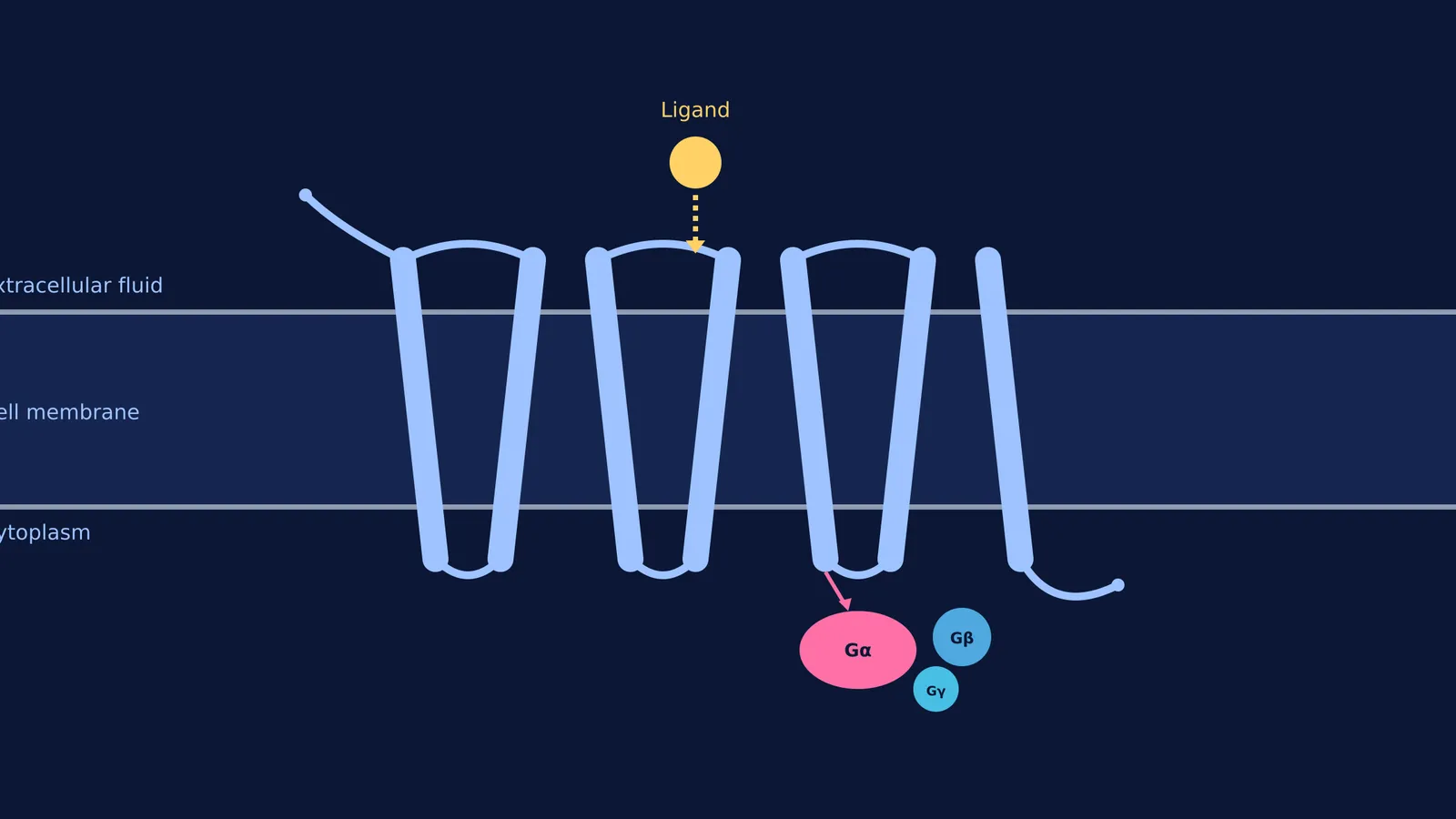
In the 1980s, Brian Kobilka joined the research and took the next massive step: he isolated the gene that coded for this receptor. When he analysed the gene, he found a sequence coding for seven long, hydrophobic (water-repelling) spirals. This meant the protein threaded itself back and forth through the lipid bilayer of the cell membrane exactly seven times (The Nobel Prize in Chemistry 2012).
Kobilka realised this seven-helix structure was identical to rhodopsin, a light-sensitive receptor in the eye. They had not just found one receptor; they had discovered an entire family of proteins that work the same way, now known as seven-transmembrane (7TM) receptors or GPCRs.
Much like the work detailed in Brown, Goldstein and the discovery of the LDL receptor, mapping these structures transformed biology from a science of observation into one of precise molecular mechanisms.
How do G protein-coupled receptors work?
A GPCR works like a lock and key that triggers a domino effect inside the cell.
The signal arrives: A molecule (the ligand) binds to the receptor on the outside of the cell membrane.
The shape changes: The binding causes the entire seven-helix protein to twist and change its three-dimensional shape.
The G-protein activates: This shape change on the inside of the cell wakes up an attached G-protein complex. The G-protein splits apart, and its subunits travel through the cell interior to trigger enzymes and initiate the final biological response.
In 2011, Kobilka's team achieved what many considered impossible: they used X-ray crystallography to capture an image of the β-adrenergic receptor at the exact moment it transferred a signal from a hormone on the outside to a G-protein on the inside.
If a student is struggling to picture how a 3D protein threads through a membrane and changes shape, Fermi's AI tutor can help. Fermi teaches — it holds a line of reasoning across a whole session, notices where a student is stuck, and decides whether to explain, ask, or show, writing on a board and drawing the figure as it goes.
How many GPCRs are in the human body?
When the human genome was mapped, researchers found that close to 1,000 genes code for GPCRs (The Nobel Prize in Chemistry 2012).
These receptors govern nearly every physiological process. Around 500 of these genes are dedicated to the olfactory system, allowing us to distinguish thousands of different smells. Another 333 receptors detect hormones and neurotransmitters like dopamine, serotonin, and histamine. They are the reason you can see light, taste food, and maintain balance. They are also central to physiological reflexes, such as those explained in How does the body know when to feel thirsty?
Because GPCRs are the primary way cells interact with their environment, they are incredibly important in medicine. According to the Nobel committee, around 50% of all medications act through these receptors. This includes beta-blockers for heart conditions, antihistamines for allergies, and many psychiatric drugs.
For a broad overview of how these receptors fit into organ systems, the Fermi Concept Library contains every concept in physics, chemistry, biology, mathematics and computing, each with its own lesson and its place in the map.
How to study cell signaling for biology exams?
Memorising dozens of pathways for exams like NEET-UG or class 12 boards can feel overwhelming. Instead of learning them as disconnected facts, use a four-part framework for every signaling pathway:
The Ligand: What is the messenger? (e.g., adrenaline).
The Receptor: What does it bind to? (e.g., β-adrenergic receptor, which is a GPCR).
The Transducer: How does the signal cross the membrane? (e.g., the G-protein activates and triggers an enzyme).
The Effector: What is the final biological result? (e.g., the cell breaks down glycogen to release glucose).
By breaking pathways down into these four logical steps, you build a mental map that is easier to remember and apply to complex exam questions.