On this page
Before the 1950s, biologists who wanted to look inside a cell with an electron microscope had a cutting problem. The microscope could show tiny detail, but only if the sample was a slice thin enough for electrons to pass through. A Venezuelan doctor, Humberto Fernández-Morán, solved a big part of that problem with a blade made of diamond.
Who was Humberto Fernández-Morán?
He was born in Maracaibo, Venezuela, on 18 February 1924 and died in Stockholm on 17 March 1999, according to his Wikipedia biography, which also records a John Scott Award in 1967 for the diamond knife. The US National Library of Medicine's guide to his papers calls him a physician and biophysical researcher. It says he earned his medical degree in Munich in 1944, founded the Venezuelan Institute for Neurological and Brain Studies, and moved to the University of Chicago in 1958. His career centred on electron microscopy, cryomicroscopy and ultramicrotomes.
Why does an electron microscope need such thin slices?
A transmission electron microscope sends a beam of electrons through the sample, so the sample has to let the beam through. The Wikipedia article on ultramicrotomy, citing the textbook by Bozzola and Russell, notes that electrons accelerated at 50 to 125 kV cannot pass through biological material much thicker than 150 nm, and that sections of 30 to 60 nm give the best resolution.
The Michigan Tech electron microscopy lab describes its ultramicrotome as cutting sections typically 50 to 100 nanometres thick.
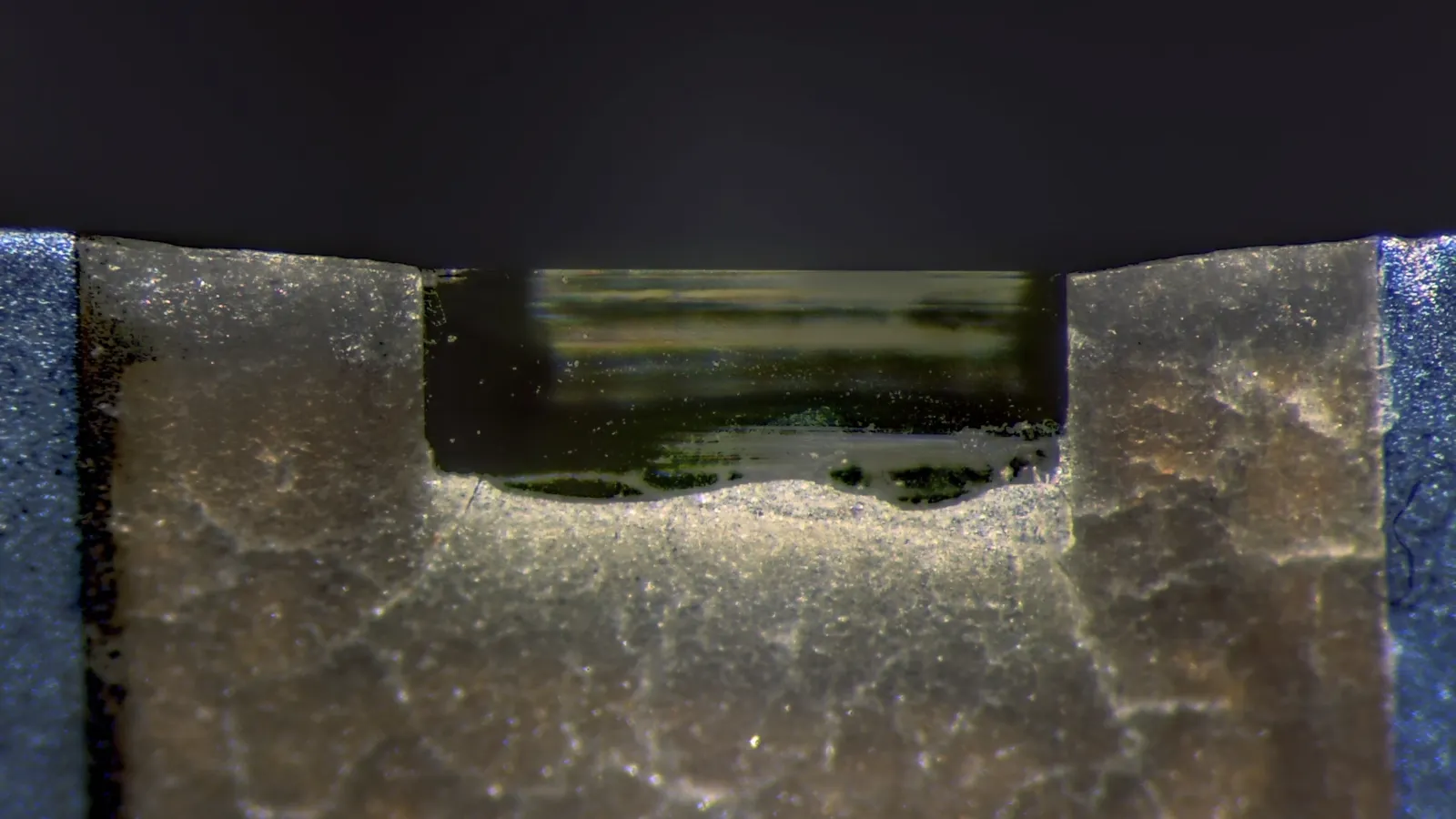
Glass came first. In 1950, Latta and Hartmann found that the edge of broken glass could cut thin sections for electron microscopy, as the Wikipedia article on the diamond knife explains. Glass dulls quickly, though: the same article says a skilled operator gets a small number of ultrathin sections from a glass knife, but thousands from one area of a diamond knife.
How does a diamond knife cut ultra-thin sections?
Fernández-Morán published "A diamond knife for ultrathin sectioning" in Experimental Cell Research in 195390112-8). In 1956 he followed it with a paper on using the knife on biological tissues and metals, written at the Venezuelan Institute for Neurology and Brain Research in Caracas.
His US patent, US3060781, gives the details. Its priority date is 4 September 1954 and it was published on 30 October 1962.
The edge: a diamond cutting edge 0.001 to 0.01 micron thick.
The stone: industrial diamonds such as boarts, which are built up of very thin layers and split cleanly along them.
The polish: grinding and polishing with very fine diamond powder on a disc spinning at tens of thousands of revolutions per minute.
The slices: sections of the order of 0.001 to 10 microns thick, from both soft and hard materials.
The patent calls the hardness of diamond unsurpassed, so its edge stays sharp far longer than glass. The diamond knife article adds that the edge is free of imperfections, which helps it cut sections of very regular thickness.
Worked example: how thin is a 70 nm slice, really?
Try the arithmetic yourself. It is good practice for unit conversion.
Pick a slice thickness inside the 50 to 100 nm lab range above: say 70 nm.
Convert a sample block 1 mm deep into nanometres: 1 mm = 1,000 micron = 1,000,000 nm.
Divide: 1,000,000 ÷ 70 ≈ 14,300 slices from one millimetre of tissue.
Compare with the edge: a 70 nm slice is 7 to 70 times thicker than an edge of 1 to 10 nm.
Sense check: the ultramicrotomy article above says its best-resolution sections are like splitting a 0.1 mm human hair into 2,000 slices. Check it: 0.1 mm ÷ 2,000 = 50 nm.
Step 3 shows why a lasting edge mattered. A blade that dulls after a few cuts cannot work through a block like that; a diamond edge can.
If conversions like micron to nanometre trip you up, the Fermi Problem Library has worked problems with a step-by-step solver that explains the reasoning rather than printing an answer.
How can you use this story in biology class?
School biology often asks why electron microscopes see more than light microscopes. The usual answer is resolution, which our explainer on why an electron microscope has better resolution covers. Fernández-Morán's story adds the step people skip: the sample has to be made thin enough first.
When you revise, check that you can explain these three points:
Electrons, not light, form the image, so the sample must be very thin.
Ultrathin sections are about 50 to 100 nm, cut on an ultramicrotome.
Glass knives came first; diamond knives last much longer and cut evenly.
To see where the microscope itself came from, read about Ernst Ruska and the first electron microscope. For what thin sections went on to reveal inside cells, try George Palade and the ribosomes in the cell's protein factory.
If ideas like resolution or wavelength still feel shaky, the Fermi Concept Library covers every concept in physics, chemistry, biology, mathematics and computing, each with its own lesson and its place in the map.