

On this page
Why can we see a whole cell under a light microscope but need an electron microscope to see the viruses inside it or the atoms in a crystal? It all comes down to wave-particle duality and the fundamental limits of diffraction.
Why does an electron microscope have better resolution?
Whenever a wave hits an obstacle or passes through an opening, it bends. This phenomenon, called diffraction, sets a hard limit on what any microscope can resolve. A microscope cannot distinguish two points that are closer together than roughly half the wavelength of the beam being used to look at them.
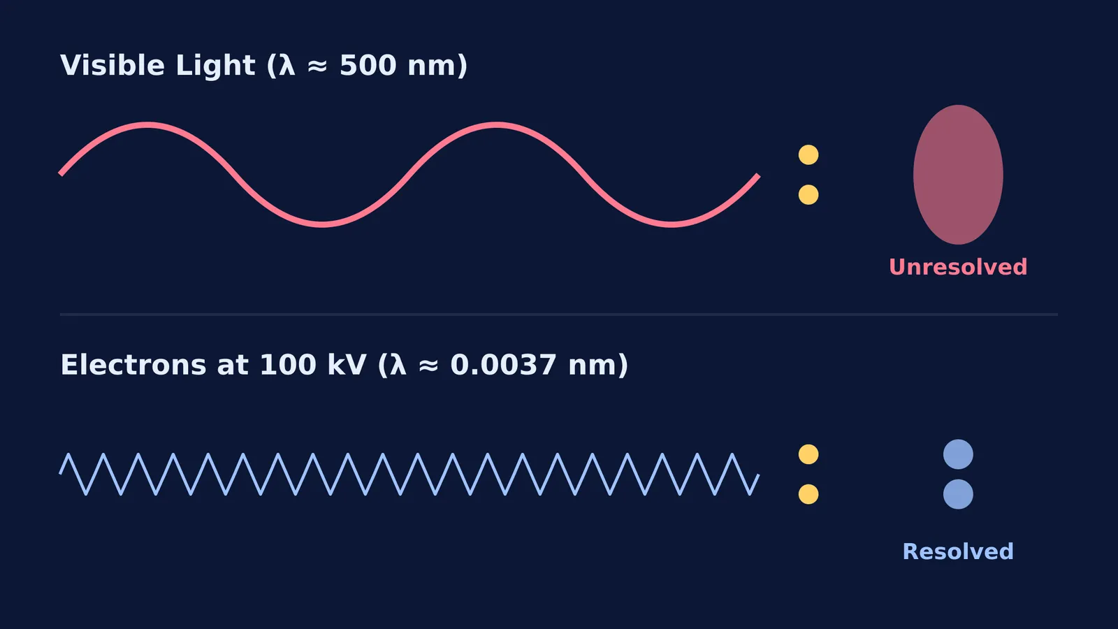
Visible light has wavelengths ranging from about 380 to 750 nanometers (Wikipedia). Because of this, even a perfectly engineered optical microscope with flawless glass lenses is physically limited to a resolution of about 200 nm (Wikipedia). If two structures are 50 nm apart, the light waves simply wash over them, blurring them into a single, unresolved blob.
To see smaller details, you need a much shorter wavelength. By replacing light with a beam of fast-moving electrons, scientists bypass the limitations of the visible spectrum and shatter the traditional diffraction limit.
How do you calculate the wavelength of an electron?
According to the de Broglie relation, moving particles like electrons behave as waves, and their wavelength (λ) depends on their momentum (p):
λ = h / p
where h is the Planck constant (Wikipedia). By accelerating electrons through a high voltage, we give them a massive momentum and, consequently, an incredibly short wavelength.
This wavelength is more than 100,000 times shorter than that of visible light (Wikipedia). The mathematics behind wave-particle duality can feel abstract when you first encounter it. If you are struggling with these calculations, the Fermi Concept Library can help you trace exactly how the mass, charge and accelerating potential interact to change the final matter wave.
What is the maximum resolution of an electron microscope?
Because of their tiny wavelength, electron microscopes have a much higher resolution of about 0.1 nm, allowing scientists to see viruses, proteins, and large molecules clearly (Wikipedia).
In advanced high-resolution transmission electron microscopy (HRTEM), hardware correctors push this resolution even further—to below 0.05 nm, or 50 picometres. At this scale, it is possible to map the positions of individual atoms in a crystal lattice. However, practical resolution is rarely perfect. It is often limited by spherical aberration in the magnetic lenses used to focus the electron beam, and by the preparation of the biological samples themselves.
If you ever find yourself struggling to visualise how waves interact with gaps and edges, or why short wavelengths maintain their path better without spreading out, the Fermi Simulation Lab lets you adjust wavelengths and slit sizes to see diffraction patterns emerge in real time.
How do transmission and scanning electron microscopes differ?
While all electron microscopes rely on short de Broglie wavelengths, they apply this principle in different ways to capture different types of images.
In a Transmission Electron Microscope (TEM), the electron beam passes directly through an ultra-thin sample. This is much like how light passes through a glass slide in an optical microscope. The TEM provides the highest possible resolution, making it the tool of choice for looking at internal cellular structures or atomic arrangements.
In contrast, a Scanning Electron Microscope (SEM) bounces a focused beam of electrons off the surface of a sample. As the beam scans across the object, it knocks loose secondary electrons, which are collected to build a detailed, three-dimensional topographical map. Because it relies on surface interactions rather than passing entirely through the material, an SEM typically has a slightly lower resolution (around 1 nm) than a TEM (Wikipedia). However, it excels at showing the outer shapes of insects, pollen grains, and fractured metals.
Understanding resolution is a key part of bridging the number of photons emitted per second in classical optics with the strange quantum reality that rules the nanoscale. Whether you are tracking displacement and pressure waves in sound or the probability waves of electrons, the rule remains consistent: shorter waves reveal smaller worlds.


