On this page
Imagine someone who talks easily, yet makes little sense and cannot follow what you say back. In 1874 a 26-year-old German doctor named Carl Wernicke argued that this pattern comes from damage to one part of the left temporal lobe. That region now carries his name.
Who was Carl Wernicke?
Wernicke was born on 15 May 1848 in Tarnowitz, Upper Silesia, and trained in medicine at the University of Breslau, according to his Wikipedia biography. He worked at Breslau's All Saints Hospital and spent six months in Vienna with the neuroanatomist Theodor Meynert. In 1904 he took charge of the psychiatry and neurology clinic at Halle. He died in June 1905 after a cycling accident in the Thuringian Forest.
What did Wernicke discover in 1874?
Wernicke's monograph, *Der aphasische Symptomencomplex* ("The aphasia symptom complex"), appeared in 1874, soon after his time in Vienna. A 2024 historical review in World Neurosurgery describes it as the first description of a sensory speech centre and of sensory aphasia. A 150th-anniversary analysis by Ardi Roelofs in Brain Structure and Function counts ten patients in the book, four of them with autopsy results.
Susanne Rother, aged 75, had a stroke and afterwards spoke fluently, but she mixed up words and had trouble understanding them. Her autopsy showed damage to the left superior temporal gyrus, Roelofs reports. Another patient, Rosina Peter, had almost the opposite problem. At first she produced only an unintelligible mumble and later became completely speechless, yet her understanding was spared. Her autopsy found damage to the white matter beneath Broca's area.
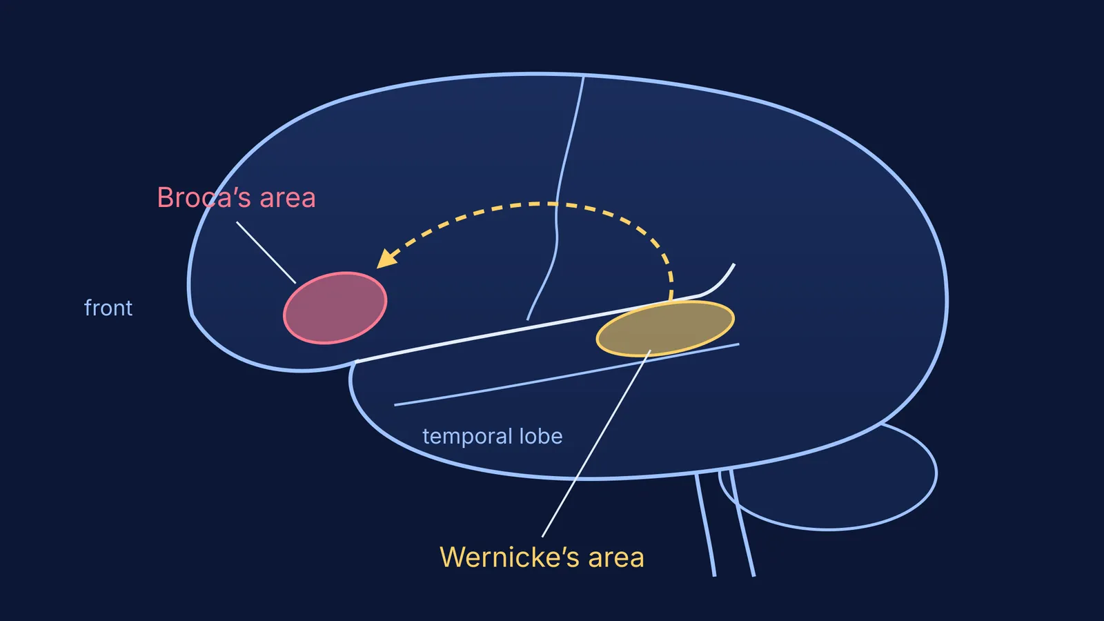
Wernicke's model, as Roelofs translates it, put the sound images of words in the superior temporal gyrus, because that area is sensory. It put the movement images for speaking in the inferior frontal gyrus, because that area is motor, and it joined the two with association fibres. Wernicke used the model to predict what damage in different places would do. If the link were cut while both ends survived, understanding should stay intact but wrong words should creep into speech, the World Neurosurgery review explains. Wernicke called this disorder *Leitungsaphasie*, now known as conduction aphasia.
The idea built on the work of Paul Broca and the patient who could say only one word. Broca linked producing speech to the frontal lobe. Wernicke added the receiving side.
What does damage to Wernicke's area look like today?
The US National Institute on Deafness and Other Communication Disorders defines aphasia as a disorder caused by damage to the brain areas responsible for language, usually from a stroke or traumatic brain injury. People with Wernicke's aphasia speak fluently in long sentences that carry little meaning, and they have difficulty understanding language, whether spoken, written or signed. People with Broca's aphasia speak in short, effortful phrases but usually understand better. The same NIDCD page says about one third of stroke survivors develop aphasia.
The US CDC lists these signs on its stroke symptoms page and notes that the treatments that work best are available only if the stroke is diagnosed within 3 hours of the first symptoms.
Is Wernicke's area really the brain's comprehension centre?
Not on its own. In a 2015 study in Brain, M-Marsel Mesulam and colleagues studied 72 people with primary progressive aphasia, a slowly worsening language disorder. They found that losing neurons in temporoparietal areas traditionally counted as Wernicke's area left the understanding of single words intact. Severe trouble understanding single words went with shrinkage at the front of the left temporal lobe instead. The authors concluded that a single patch of cortex equally critical for understanding both words and sentences is unlikely to exist.
So the textbook picture is a useful first map, not the whole territory. Language depends on networks of connected regions, which is also what Wilder Penfield found when he mapped the brain during surgery.
How do you tell Broca's and Wernicke's areas apart in an exam?
Use this three-question check on any case description:
Is speech fluent? Fluent but meaningless points to Wernicke. Slow and effortful points to Broca.
Is understanding damaged? Poor understanding points to Wernicke. Fairly good understanding points to Broca.
Which lobe? Wernicke's area is in the temporal lobe, at the back of the superior temporal gyrus. Broca's area is in the frontal lobe, in the inferior frontal gyrus.
Worked example: "A patient chats readily but answers 'What is your name?' with an unrelated, jumbled sentence." Speech is fluent (yes), understanding is damaged (yes), so the answer is the temporal lobe, Wernicke's area.
For the bigger layout, our guide to the parts of the human brain and their functions covers the forebrain, midbrain and hindbrain. If a related idea such as the reflex arc or the synapse is still shaky, the Fermi Concept Library has a lesson for every concept in biology and shows its place in the map of related ideas.