

On this page
Try reading this sentence while looking an inch to the right of your screen. The words become a blur, and you can no longer make out individual letters. Why do you have to look straight at a book to read it? The answer lies in the physics of the eye and a highly specialised patch of the retina called the fovea centralis.
What is the macula lutea?
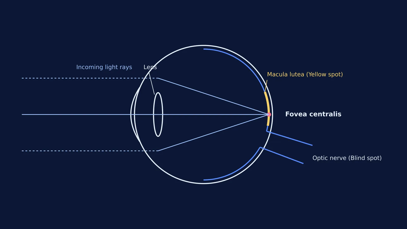
When light enters your eye, the cornea and the lens act much like refraction through a glass sphere, focusing the incoming light rays onto the back inner wall of the eyeball, which is known as the retina. Directly opposite the lens, near the centre of the retina, is an oval area about 5.5 millimetres wide known as the macula lutea (Wikipedia).
In Latin, _macula lutea_ literally translates to "yellow spot". It gets this distinctive yellow colour from a high concentration of dietary pigments, specifically lutein and zeaxanthin (Hong Kong Journal of Ophthalmology, 2000). These pigments are crucial for eye health because they act as internal natural sunglasses. They absorb harmful, high-energy blue and ultraviolet light before those rays can reach and damage the delicate, highly sensitive light-detecting cells lying beneath the surface.
Why is vision sharpest at the fovea centralis?
At the exact centre of the macula lutea lies a very shallow depression or pit called the fovea centralis. This tiny pit acts as the absolute "bullseye" of your visual field.
The human retina contains two main types of photoreceptor cells: rods, which are highly sensitive and detect dim light and shadows; and cones, which require brighter light to function but detect rich colour and fine spatial detail. Vision is at its sharpest at the fovea because it contains the highest density of cone cells found anywhere in the entire eye. A landmark anatomical study found that the peak cone density in the human fovea averages 199,000 cones per square millimetre (Curcio et al., 1990).
In the very central region of the fovea (often called the foveola), there are exactly zero rods. This rod-free zone is about 0.35 millimetres wide, which corresponds to just 1.25 degrees of your visual field. This narrow angle explains exactly why you must point your eyes directly at the specific thing you want to see clearly: you are physically aligning the incoming light rays so they strike this tiny, 1.25-degree patch of tightly packed cones.
To make the resulting image even sharper, the fovea has evolved a unique structure. It has no blood vessels covering its surface, and the upper layers of retinal nerve cells are physically pushed to the side, forming the pit. This allows incoming light to hit the cone cells directly without any scattering. Furthermore, each cone cell in the fovea connects to a single, dedicated optic nerve fibre, sending a pure, unmixed "pixel" of visual data straight to the visual cortex.
How does the fovea differ from the blind spot?
While the fovea centralis is densely packed with photoreceptors to provide maximum visual clarity, the blind spot has no photoreceptor cells at all.
Also known scientifically as the optic disc, the blind spot is located slightly to the nasal side of the macula (towards your nose). This circular area acts as the main exit door of the eye. It is the precise location where all the individual nerve fibres from across the entire retina bundle together to form the thick optic nerve and begin their journey to the brain (you can review how the brain processes these complex visual signals in our comprehensive guide to the parts of the human brain). Because this exit space is completely taken up by exiting nerve fibres and entering blood vessels, there is simply no physical room left for any rods or cones (Wikipedia).
Any light rays that happen to fall onto the optic disc cannot be detected by the eye. We do not normally notice this permanent hole in our visual field because our brain is incredibly good at seamlessly filling in the missing details, using matching background patterns and overlapping data provided by the other eye.
Preparing for exams
When studying the anatomy of the eye for Class 10 science or NEET biology, keeping the roles of the yellow spot and the blind spot distinct is a frequent exam requirement. Remember that the macula is directly opposite the lens for sharp central vision, while the optic disc is the blind exit below it.
In the Concept Library, you can explore the structure of the human eye and other sensory organs interactively. Fermi's AI tutor works through the diagrams with you, checking your understanding of where images form and why, rather than just giving you a textbook page to memorise.


