On this page
Walk out of a dark cinema hall into a sunny afternoon and everything is too bright for a moment. Then it settles, partly because your pupils shrank without you deciding anything. Here are the two muscles that do it, the nerve route that triggers it, and how to write it up in an exam.
What do the circular and radial iris muscles do?
The pupil is not a structure of its own. It is the hole in the middle of the iris, the coloured ring of your eye, and the iris contains two sets of smooth muscle that pull in opposite directions.
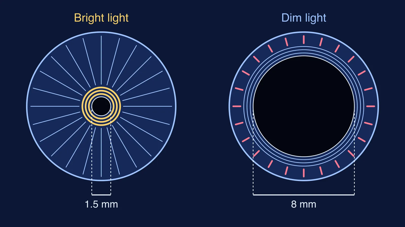
Sphincter pupillae (circular muscle): an annular band of smooth muscle encircling the pupil. When it contracts, the ring tightens and the pupil gets smaller.
Dilator pupillae (radial muscle): radially oriented smooth muscle fibres running out from the pupil like spokes. When they contract, they pull the edge of the pupil outward and the pupil gets bigger.
Both descriptions come from a review of how the nervous system controls the eye by David McDougal and Paul Gamlin in Comprehensive Physiology. Their abstract also gives the wiring: ciliary ganglion neurons (parasympathetic) supply the sphincter pupillae to constrict the pupil, and superior cervical ganglion neurons (sympathetic) supply the dilator pupillae to widen it.
If the sympathetic and parasympathetic labels are hazy, our guide to nervous system divisions: CNS, PNS, somatic and autonomic lays them out.
Why does the pupil get smaller in bright light?
Like the knee jerk reflex, it runs on a fixed loop you cannot switch off. The pathway, as summarised in Wikipedia's pupillary light reflex entry, which cites the textbook Adler's Physiology of the Eye, runs:
Light falls on the retina and excites retinal ganglion cells.
The signal travels along the optic nerve to the pretectal nucleus in the midbrain.
The pretectal nucleus signals the Edinger-Westphal nucleus on both sides.
Parasympathetic fibres run in the oculomotor nerve to the ciliary ganglion.
The ciliary ganglion fires the sphincter pupillae, which contracts and narrows the pupil.
In dim light the opposite happens: the sphincter relaxes and the sympathetic supply to the radial dilator widens the pupil, letting in more light so you can still see.
The reflex is fast. McDougal and Gamlin report that the pupil constricts with a latency of about 100 ms, with peak constriction about 300 to 500 ms after the light arrives (review). The same review notes that in humans the direct reflex (the lit eye) and the consensual reflex (the other eye) are essentially equal, which is why shining light in one eye shrinks both pupils.
How much light does a smaller pupil actually block?
The normal human pupil can change diameter from 8 mm to 1.5 mm, which corresponds to roughly a 28-fold change in area (McDougal and Gamlin). You can check it yourself.
The light the pupil admits is proportional to its area, and area goes as the square of diameter:
Wide pupil: area = π × (4 mm)² ≈ 50.3 mm²
Narrow pupil: area = π × (0.75 mm)² ≈ 1.77 mm²
Ratio: 50.3 ÷ 1.77 ≈ 28
So halving the diameter cuts the light by four times, not two. A camera's aperture follows the same square law, and the review describes the pupil as an aperture stop that controls the amount of light striking the retina.
Can the pupil reflex protect your eyes from the Sun?
Only partly. The reflex handles everyday bright light, but it cannot make staring at the Sun safe. The American Academy of Ophthalmology's EyeWiki page on solar retinopathy describes it as a photochemical injury to retinal tissue, usually at the fovea, and says prevention means not looking directly at the Sun or an eclipse at all.
How do you write the pupil reflex answer in an exam?
For "Why does the pupil become small in bright light?", cover these points:
Name the reflex: the pupillary light reflex, an involuntary response.
Name the muscle: the circular sphincter pupillae contracts.
Name the opposing muscle: the radial dilator pupillae, which widens the pupil in dim light.
Give the purpose: less light reaches the retina in bright conditions.
Add the nerve route for longer answers: retina, optic nerve, midbrain, oculomotor nerve, iris.
Then see it yourself. Sit facing a mirror in a dim room for a minute, switch the light on, and watch your pupils shrink. Next, cover one eye with your palm and uncover it while watching the other pupil. Afterwards, try the card test that finds your blind spot.
If the autonomic wiring still feels shaky, the Fermi Concept Library gives every biology and physics concept its own lesson and its place in the map. For numericals, the Fermi Problem Library has worked problems with a step-by-step solver that explains the reasoning rather than printing an answer.