On this page
An ultrasound scanner is an echo-timing machine. The probe, called a transducer, both sends out ultrasound and picks up the echoes that bounce back, as the US National Institute of Biomedical Imaging and Bioengineering (NIBIB) explains. Ultrasound means sound above 20 kHz, beyond the range of human hearing, and the same NIBIB page notes that most scanners work at much higher frequencies, in the megahertz range.
How does an ultrasound scan make a picture?
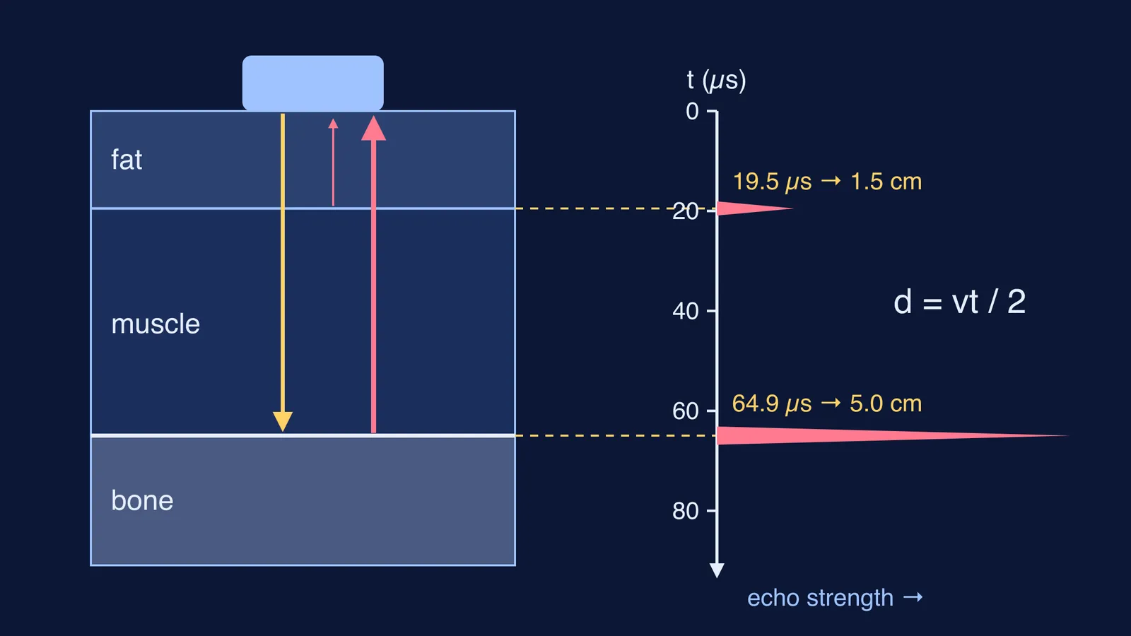
The probe sends a short pulse, then listens. NIBIB says the waves are reflected back by boundaries between tissues, such as fluid and soft tissue or tissue and bone. In NIBIB's words, "Using the speed of sound and the time of each echo's return, the scanner calculates the distance from the transducer to the tissue boundary." Those distances are then used to build a two-dimensional image. OpenStax adds that the beam is swept across the area and the image maps the intensity of the reflected sound, so stronger echoes show up brighter.
For the history, read how Ian Donald made the first ultrasound scans of babies in the womb.
Why do echoes come from boundaries between tissues?
Sound reflects wherever the medium changes. Physicists measure how much a medium resists sound with acoustic impedance, Z, which is density multiplied by the speed of sound in that material (OpenStax College Physics 2e, section 17.7). The bigger the jump in Z at a boundary, the stronger the echo. The same OpenStax section gives the fraction of sound intensity reflected as (Z2 − Z1)² ÷ (Z1 + Z2)².
We put OpenStax's impedance table into that formula:
Fat to muscle: only 1.4% of the intensity is reflected (the textbook's worked example), so most sound carries on and the scan sees layer after layer.
Muscle to bone: bone (5.7 to 7.8 × 10⁶ kg/(m²·s)) is far above muscle (1.70 × 10⁶), so about 29% to 41% bounces back and bone looks bright.
Tissue to air: air is just 429 kg/(m²·s), so about 99.9% is reflected and almost nothing gets through.
That is why the technician spreads gel on your skin first: NIBIB says it stops air pockets forming between probe and skin, which would block the waves.
How does echo time turn into depth?
This is the echo formula from the school sound chapter, used inside a hospital. The sound goes down to the boundary and comes back, so it covers twice the depth: 2d = v × t, which means d = vt ÷ 2. Scanners assume one average speed for soft tissue, and OpenStax gives the speed of sound in tissue as about 1540 m/s.
Worked example: an echo returns 65 microseconds (µs) after the pulse leaves the probe.
Write the round trip: 2d = v × t.
Put in the values: 2d = 1540 m/s × 65 × 10⁻⁶ s = 0.100 m.
Halve it: d = 0.050 m, so the boundary is 5.0 cm below the probe.
A handy rule follows: each centimetre of depth adds about 13 µs to the echo time (2 × 0.01 m ÷ 1540 m/s). Even a boundary 10 cm deep answers in about 130 µs, so one second has room for thousands of send-and-listen cycles.
Why not always use the highest frequency?
Higher frequency means shorter wavelength, and OpenStax points out that you cannot see details much smaller than the wavelength of your probe. At 7 MHz the wavelength in tissue is 1540 ÷ 7,000,000 ≈ 0.22 mm. The catch is that high frequencies fade faster: OpenStax gives a rule of thumb that you can effectively scan to a depth of about 500 wavelengths, roughly 11 cm at 7 MHz. Halve the frequency to 3.5 MHz and the wavelength doubles to 0.44 mm, so by the same rule the reach doubles to about 22 cm while detail gets coarser. Detail or depth: that trade-off sets a probe's frequency.
The US Food and Drug Administration says ultrasound uses non-ionising radiation and has an excellent safety record, but it can heat tissue slightly, so it discourages scans for non-medical reasons such as keepsake videos. Whether you need a scan, and what it shows, is for your doctor to decide.
How does this connect to Class 9 sound?
Ultrasound sits in the school sound chapter next to echoes, SONAR and the range of human hearing. Our guide to sound in Class 9 covers the whole chapter, and the uses of infrasonic and ultrasonic sound lists other uses. Check yourself:
Can you say why scanners use ultrasound rather than audible sound? (Shorter wavelength, finer detail.)
Can you write 2d = vt and explain where the 2 comes from?
Can you explain why echoes come from boundaries, not from the middle of one uniform tissue?
Can you explain why the gel is needed?
Can you redo the 65 µs example without notes, then try an echo at 26 µs? (Answer: 2.0 cm.)
Pulses and echoes are easier to picture when you watch them move. The Fermi Simulation Lab has interactive simulations for ideas that are easier to see than to read, and the Fermi Concept Library gives each physics concept, such as reflection of sound, its own lesson.