
On this page
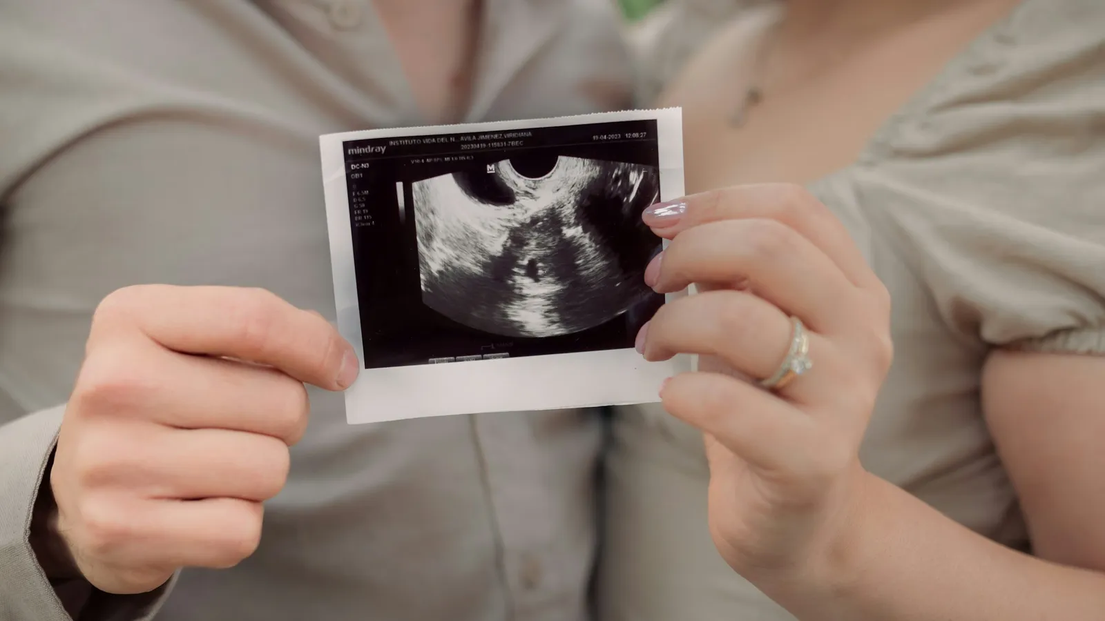
For many people, the first picture ever taken of them is a grey, grainy ultrasound scan from before they were born. That scan exists because a Glasgow obstetrician in the 1950s looked at a machine used to find cracks in steel and wondered whether it could find problems inside a patient. This post covers who Ian Donald was, how his team built the first obstetric scans, the physics of echo timing with a worked example, and what you can take from his way of working.
Who was Ian Donald?
The Embryo Project Encyclopedia at Arizona State University records that Donald was born on 27 December 1910 in Cornwall, England, and died in 1987. He went to school in Scotland and South Africa, took an arts degree at the University of Cape Town and then studied medicine at St Thomas's Hospital in London.
During the Second World War he served as a medical officer in the Royal Air Force, where, the encyclopedia notes, he developed a keen interest in sonar and radar. In September 1954 he became Regius Professor of Midwifery at the University of Glasgow.
How did a factory machine become a medical scanner?
In spring 1955 one of Donald's patients introduced him to her husband, a director of the boilermaking firm Babcock and Wilcox. Their technician Bernard Donnelly showed him an ultrasonic flaw detector, a machine that sends short pulses of high-frequency sound into metal and listens for echoes from hidden cracks. He tested it by bouncing the beam off the bone in his own thumb.
In late 1956 he met Tom Brown, a 23-year-old engineer at the Glasgow firm Kelvin and Hughes, who was already working with physician John MacVicar. In 1957 the team scanned a woman who had been diagnosed with inoperable stomach cancer. The echoes pointed to a large ovarian cyst instead. Surgery confirmed it, and the cyst was removed.
In June 1958 Donald, MacVicar and Brown published Investigation of abdominal masses by pulsed ultrasound in The Lancet. Historian Malcolm Nicolson, in a lecture for the Royal College of Physicians of Edinburgh, notes that this paper contained the first ultrasound images of the fetus ever published.
How does ultrasound imaging work?
The US National Institute of Biomedical Imaging and Bioengineering explains that ultrasound means sound above about 20 kHz, beyond human hearing, and that medical probes work in the megahertz range. The probe uses piezoelectric crystals, which turn an electric signal into sound and returning echoes back into electric signals.
The idea is the same as sonar: send a pulse, time the echo, and work out the distance. Each boundary between tissues reflects part of the sound, and the time delay tells the machine how deep that boundary is.
Worked example: echo depth and wavelength
The NCERT Class 11 Physics chapter on Waves has an exercise on a hospital scanner in tissue where sound travels at 1.7 km/s and the frequency is 4.2 MHz. We use those numbers.
Wavelength. λ = v / f = 1,700 / (4.2 × 10⁶) = 4.05 × 10⁻⁴ m, about 0.4 mm.
Why this matters. Fine detail needs short wavelengths, which is why medical probes use megahertz frequencies.
Echo depth (sample number). Suppose an echo returns 100 μs after the pulse. Depth = v × t / 2 = 1,700 × (100 × 10⁻⁶) / 2 = 0.085 m, or 8.5 cm.
Check the reverse. A boundary 8.5 cm deep means a round trip of 17 cm. Time = 0.17 / 1,700 = 1.0 × 10⁻⁴ s, which is 100 μs.
What mistakes do students make with echo problems?
Forgetting the round trip. Using d = vt without dividing by two doubles the depth.
Unit slips. Microseconds are 10⁻⁶ s and megahertz are 10⁶ Hz. Write the powers of ten out every time.
Using the speed in air. Sound is far faster in tissue and water than in air. Use the speed given for the medium.
Mixing up frequency and wavelength. When sound enters a new medium its frequency stays the same; its speed and wavelength change.
Our post on sound for Class 9 covers the basics of echoes, and the story of sonar itself is in Paul Langevin and ultrasound under water.
If the link between pulse, echo and depth is hard to picture, the Fermi Simulation Lab has interactive simulations for ideas that are easier to see than read.
What can a student learn from Donald's way of working?
Borrow tools from other fields. A machine built for boilers became one of medicine's most used tests.
Work with people who know what you don't. Donald knew patients; Brown knew electronics. Neither could have done it alone.
Keep judgement in charge of the machine. Nicolson quotes Donald warning that "the days of push-button diagnosis are no nearer". A scan still needs someone to interpret it.
Next step: work out the echo time from a boundary 12 cm deep in tissue, using 1.7 km/s, then check your answer by reversing the calculation.