On this page
When you place an unknown plant cross-section under a microscope, distinguishing a root from a stem takes just a moment. Confirming whether that root belongs to a dicot or a monocot requires looking deeper into the central vascular cylinder, known as the stele. While both plant groups share an outer protective envelope and a wide ground cortex, their vascular architecture reflects fundamentally different growth strategies.
What does a root transverse section reveal under the microscope?
Every young angiosperm root transverse section shows a conserved radial organisation that moves inwards across four distinct zones, as documented in OpenStax Biology 2e. On the outside lies the epiblema, a single cell layer bearing unicellular root hairs that absorb soil water. Beneath it extends the cortex, composed of multiple layers of thin-walled, loosely packed parenchyma cells with visible intercellular spaces.
The cortex terminates at the endodermis, a single ring of barrel-shaped cells whose radial and transverse walls carry waxy, suberin-rich Casparian strips. This ring acts as an apoplastic checkpoint, forcing all water and dissolved minerals through cell membranes before entering the vascular core. Inside the endodermis lies the stele, bounded by the pericycle. In all roots, the vascular tissues exhibit a radial arrangement, where xylem and phloem occupy alternating radii, and an exarch xylem pattern, where smaller protoxylem vessels point outward toward the pericycle while wider metaxylem vessels face inward toward the centre.
If you are reviewing tissue organization across different plant organs, our guide to identifying plant transverse sections outlines how root steles contrast with stem vascular arrangements.
How do xylem bundle count and pith size differ between roots?
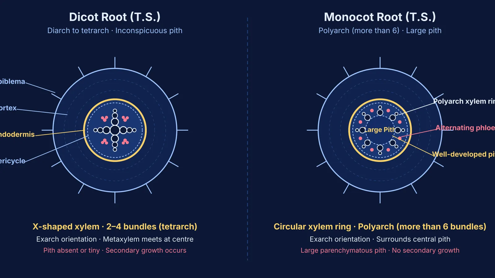
The two most reliable visual diagnostics on a prepared slide are the xylem bundle count and the proportion of central pith. According to the NCERT Class 11 Biology curriculum, a dicot root typically exhibits two to four xylem bundles—described as diarch, triarch, or tetrarch—and rarely up to six. The metaxylem vessels in a dicot root meet at the centre of the cylinder, creating an unmistakable X-shaped or star-shaped core. Because these thick-walled conducting elements fill the central space, the pith is either microscopic or entirely absent.
In contrast, a monocot root exhibits a polyarch condition, possessing more than six distinct xylem bundles arranged in an orderly circle just inside the pericycle. Rather than converging at the centre, the xylem bundles flank a spacious, well-developed central pith made of round parenchyma cells that store starch and maintain hydration.
Dicot root xylem: two to four vascular bundles forming an angular star pattern in the centre.
Monocot root xylem: more than six vascular bundles forming an open circular ring.
Dicot root pith: small, reduced, or completely inconspicuous.
Monocot root pith: large, prominent, and clearly parenchymatous.
Why does the pericycle behave differently in dicots and monocots?
Beyond static anatomy, the two root types differ in how their pericycle cells function during plant maturation. In a review in the Annals of Botany, researchers showed that root pericycle cells opposite the xylem poles retain developmental pluripotency, coordinating lateral root branching and directing the initiation of vascular cambium.
In dicot roots, the pericycle is bi-functional. It gives rise to lateral root primordia and contributes to the formation of the vascular cambium and cork cambium (phellogen). During the onset of secondary growth, parenchymatous conjunctive tissue below the phloem patches becomes meristematic, joining with pericycle cells above the protoxylem to form a continuous, undulating wavy ring. This secondary cambial cylinder later becomes circular, laying down secondary xylem toward the inside and secondary phloem toward the outside to increase root girth.
Monocot roots lack secondary growth entirely. Their pericycle produces lateral root branches but never gives rise to secondary meristems. As a result, monocot vascular bundles remain closed, and the root maintains its primary diameter throughout its lifespan. To compare how primary and lateral meristems behave during root maturation, see our overview of meristematic tissue types.
Understanding how primary cell layers transform into complex vascular organs requires seeing how developmental rules connect across chapters. The Fermi Concept Library covers every concept in biology, each with its own lesson and its place in the map, helping you explore how meristems, vascular cylinders, and secondary growth fit together.
How do you identify an unknown root slide in four steps?
When facing an unknown slide in a practical spot test, avoid jumping to conclusions based on magnification or staining colour alone. Use this systematic four-step sequence grounded in standard NCERT root anatomy criteria to verify the specimen within seconds:
Confirm the root identity: Check the outer perimeter for unicellular root hairs and verify that the xylem is exarch, with smaller vessels facing outward. This separates root slides from stem sections.
Inspect the central axis: Look straight at the center of the cylinder. A broad, light-coloured parenchymatous pith indicates a monocot, whereas a dense, dark core of meeting vessels indicates a dicot.
Count the xylem poles: Count the xylem patches along the pericycle ring. If you count two, three, or four arms in an X shape, identify it as a dicot root. If you count eight, ten, or twelve bundles in a ring, identify it as a monocot root.
Check for secondary changes: Look for a continuous wavy cambial ring or secondary wood layers between xylem and phloem patches. Any secondary thickening confirms a maturing dicot root.
For students practicing slide interpretation alongside cell division studies, our breakdown of the onion root tip practical illustrates how meristematic zones appear under identical lab conditions.
Working through realistic slide-identification scenarios builds lasting recognition. The Fermi Problem Library has worked problems with a step-by-step solver that explains the reasoning rather than printing an answer, helping you practice identifying unknown plant cross-sections.