On this page
Chlamydomonas is a single-celled green alga that frequently appears in biology exams and standard laboratory diagrams. As a model organism, it strips eukaryotic cell structure down to its absolute essentials, making it an excellent starting point for learning about plant biology, photosynthesis, and cellular movement.
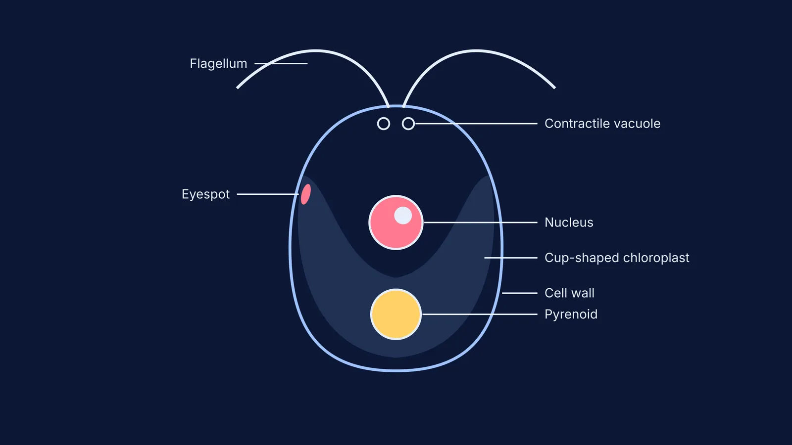
If you are asked to draw it or identify its parts in a practical exam, four distinct features set it apart from other algae and higher plants: its two flagella, a massive cup-shaped chloroplast, a starch-storing pyrenoid, and a light-sensing eyespot.
What is the structure of Chlamydomonas?
A mature *Chlamydomonas* cell is roughly spherical or pear-shaped (pyriform), with a slightly narrower front end. Unlike land plants, which rely heavily on pure cellulose for structural support, its cell wall is primarily made of hydroxyproline-rich glycoproteins.
Inside the cell boundary, the most prominent feature is the single, massive chloroplast that largely dictates the shape of the cell's interior. Above this photosynthetic organelle sits the nucleus, suspending in the cytoplasm. Near the base of the flagella, two contractile vacuoles rhythmically pump out excess water, constantly maintaining the cell's internal osmotic pressure.
Why is the chloroplast cup-shaped?
In *Chlamydomonas*, a single, large, cup-shaped chloroplast dominates the interior and defines the organism's layout (NCERT, 2024).
The "cup" or "bowl" shape effectively wraps around the central cytoplasm and cradles the nucleus. This wide, continuous structure provides a large surface area of thylakoid membranes to harvest light. For a free-floating aquatic organism, maximizing light capture from varying angles is critical for survival. Drawing the cup-shaped chloroplast first helps position all the other organelles correctly in relation to one another.
If you have concept gaps about the different structural adaptations in green algae—such as why *Spirogyra* uses a spiral ribbon while *Chlamydomonas* uses a cup—the Fermi Concept Library explains every concept in biology, each with its own lesson, to help you master these variations.
What does the pyrenoid do?
At the dense, bottom base of the cup-shaped chloroplast lies the pyrenoid.
The pyrenoid is a highly specialized, liquid-like microcompartment packed with the carbon-fixing enzyme Rubisco (Meyer et al., 2020). Its main biological job is to concentrate carbon dioxide directly around Rubisco, an adaptation known as the Carbon Concentrating Mechanism. This makes photosynthesis highly efficient even when dissolved carbon dioxide levels in the water are low.
Often, a sheath of rigid starch plates surrounds the pyrenoid, making it a highly visible circular object in both electron micrographs and student sketches. If you have already read our guide on how to sketch and label a chloroplast for NEET biology, you will notice that the mature chloroplasts of higher land plants do not typically feature this massive, centralized pyrenoid structure.
How does the eyespot sense light?
On one side of the anterior (front) portion of the chloroplast sits the eyespot, sometimes called the stigma.
The green algal eyespot apparatus is a primitive visual system composed of carotenoid-rich lipid droplets (Kreimer, 2009). It does not form complex images like an animal eye, but rather acts like a highly sensitive reflective dish. It detects both the intensity and the specific direction of incoming light. By processing these signals, the alga can swim toward optimal light levels for photosynthesis or avoid dangerously intense sunlight, a behaviour known as phototaxis.
How do the flagella work?
To physically move towards that light source, *Chlamydomonas* relies entirely on its two anterior flagella.
Both flagella are of strictly equal length, typically measuring 10 to 12 µm in wild-type cells (Asleson & Lefebvre, 1998). They emerge side-by-side from the narrow front end of the cell. During movement, they beat in a highly coordinated, breaststroke-like motion that efficiently pulls the organism through its freshwater habitat.
This biflagellated motility sets it apart from many other microscopic organisms. To understand how cell shapes dictate their lifestyles, you can compare this active swimming with the non-motile or differently structured organisms discussed in bacteria shapes: coccus, bacillus, vibrio and spirillum.
A checklist for drawing Chlamydomonas
To secure full marks on a practical diagram, run through this checklist to ensure your sketch includes every required biological component:
An outer oval-shaped cell wall that narrows slightly at the top.
Two identical flagella emerging from the front pole.
A large, U-shaped (cup-shaped) chloroplast filling the lower half of the cell.
A distinct circular pyrenoid placed squarely at the base of the chloroplast.
A circular nucleus resting in the central hollow "cup" of the chloroplast.
A small, labelled eyespot positioned on one lateral side of the chloroplast.
Two small contractile vacuoles positioned just below the base of the flagella.