
On this page
Examiners look at two things on a cell diagram: how well you drew it and how well you labelled it. The OCR biological drawing handbook says plainly that marks are awarded for both the quality of the drawing and the labelling.
What order should you draw a cell diagram in?
Draw from the outside in and label last, so no label line has to squeeze between parts.
Read the question: plant cell, animal cell, or both? Does it name the parts to label?
Draw the outline large, in the middle of the space. The learner checklist in the OCR handbook asks for a drawing that takes up at least half the page or space available.
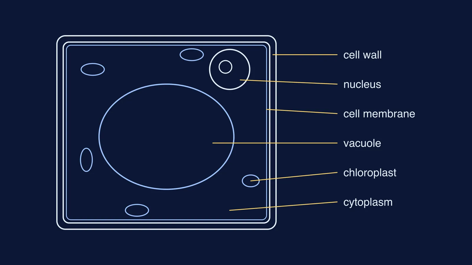
For a plant cell, draw the cell wall as the outer boundary, then the cell membrane as a separate line just inside it, which is how the NCERT textbook describes an onion cell. For an animal cell, draw a single outer membrane.
Draw the nucleus, with the nucleolus inside it as an outline.
For a plant cell, draw the large central vacuole next, then a few chloroplasts in the cytoplasm around it.
Add only the other organelles the question asks for, such as mitochondria.
Rule the label lines, write the labels, then write a title such as "Plant cell" above or below the drawing.
Unsure how the two differ? Read the cell wall versus the cell membrane.
Which labels earn marks on plant and animal cells?
The labels that score are the ones that show you know what kind of cell you drew. The NCERT Class 11 chapter Cell: The Unit of Life states the difference in one line: plant cells have cell walls, plastids and a large central vacuole, which animal cells lack, while animal cells have centrioles, which almost all plant cells lack.
Labels for both cells: cell membrane (plasma membrane), cytoplasm, nucleus, nucleolus, nuclear membrane, mitochondria.
Extra labels for a plant cell: cell wall, chloroplast, large vacuole.
Extra label for an animal cell: centriole.
If there is room and time: Golgi apparatus, endoplasmic reticulum, ribosomes, lysosome, all of which appear on NCERT's Figure 8.3.
If a term on the list is shaky, the Fermi Concept Library has a lesson for every concept in biology, so you can fix the idea before you fix the drawing.
How should pencil and label lines look?
The rules below come from the OCR handbook. It was written for UK A level students, but the habits suit most school biology drawings.
Use a sharp pencil only, not a pen or coloured pencils.
Use clear, continuous lines. A line that encloses a shape should join up neatly, without overlap.
Do not shade, stipple or cross-hatch. Draw the outline of the nucleus and leave it unshaded.
Rule every label line with a ruler, in pencil.
Start each label line exactly at the part it names, and never use arrowheads.
Keep label lines from crossing each other, and do not write on them.
Write labels horizontally. Lining them up in a column on one or both sides of the drawing is not essential, the handbook says, but it looks neater.
The same handbook warns that overlapping lines are a common error in hastily drawn sketches and are easily spotted and penalised by examiners.
What mistakes do examiners mark down?
In Cambridge's June 2024 IGCSE Biology examiner report, examiners noted that on one drawing question, of a stick insect's egg case, a significant number of candidates shaded their drawing, and reminded candidates to only draw outlines.
The same report flags other habits to avoid:
Outlines that were broken instead of continuous.
Drawings so large that they run into the printed text. The report says marks will not be awarded for size if the drawing goes into any of the text on the page.
Lines meant to be rubbed out but not fully erased.
Writing in pencil and then overwriting in pen, which makes answers hard to read.
Two more checks of our own: a plant cell where the wall and membrane have merged into one line, and label lines that stop short of their part.
A 10-minute practice plan
You need a sharp HB pencil, an eraser, a ruler and plain paper.
Minutes 0 to 3: draw a plant cell from memory, outline first.
Minutes 3 to 5: rule and write six labels: cell wall, cell membrane, nucleus, cytoplasm, chloroplast, vacuole.
Minutes 5 to 8: draw an animal cell beside it and label nucleus, cell membrane, cytoplasm, mitochondrion and centriole.
Minutes 8 to 10: check both against the rules above and circle every rule you broke.
Repeat until a round has no circles. To compare with real cells, see onion peel and cheek cell slides.
If you would rather practise with feedback, Fermi's AI tutor draws the figure and checks understanding as it goes, so you can ask why a label sits where it does.