

On this page
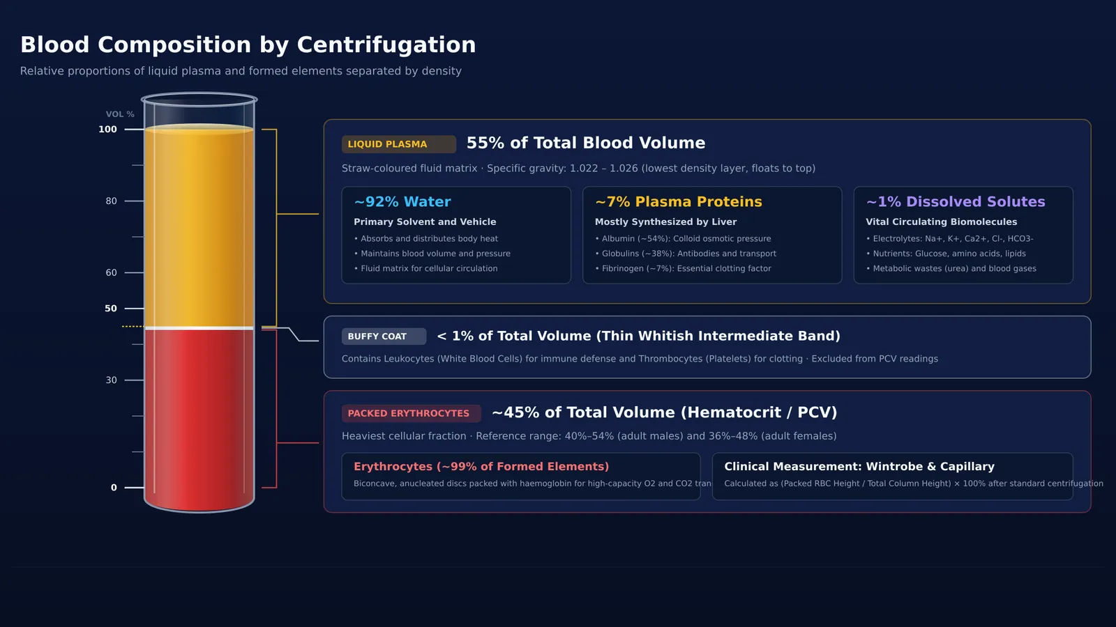
When you look at a vial of freshly drawn blood, it appears as a uniform red liquid. Yet blood is a specialised connective tissue made of living cells suspended in a liquid matrix. In human physiology, according to the NCBI StatPearls review of blood plasma physiology, whole blood divides into two principal fractions: liquid plasma, which makes up roughly 55% of total volume, and formed elements, which constitute the remaining 45%. Visualizing how these components separate under centrifugal force makes their physiological roles straightforward to master.
What is the percentage composition of blood plasma and cells?
In healthy human adults, whole blood consists of approximately 55% liquid plasma and 45% formed elements, as detailed in OpenStax Anatomy and Physiology. Plasma serves as the circulating fluid matrix, providing the volume and hydrostatic pressure required for the heart to pump nutrients, hormones, and dissolved gases throughout the body.
The 45% cellular fraction, known as the formed elements, contains three primary cell varieties:
Erythrocytes (red blood cells), making up approximately 99% of all formed elements to transport oxygen bound to haemoglobin.
Leukocytes (white blood cells), mediating immune defense against pathogens.
Thrombocytes (platelets), which are cellular fragments essential for blood clotting.
For details on cell counts and lifespans, review our guide to the formed elements of blood.
How does a centrifuge tube separate blood into layers?
In the bloodstream, continuous circulation keeps cells uniformly suspended. However, when blood is collected with an anticoagulant and spun in a centrifuge, centrifugal acceleration separates components by density.
As detailed in the NCBI StatPearls guide to hematocrit measurement, spinning anticoagulated blood in a Wintrobe tube at 3,000 revolutions per minute for 30 minutes, or in a microhematocrit capillary tube at 11,000 to 12,000 revolutions per minute for 4 to 5 minutes, yields three distinct layers:
Top layer (Plasma): Straw-coloured liquid occupying roughly 55% of the tube. Being mostly water, it has the lowest density (specific gravity of 1.022 to 1.026) and floats to the top.
Middle layer (Buffy coat): A thin, whitish layer resting above the red cells, accounting for less than 1% of total volume. It contains all leukocytes and platelets.
Bottom layer (Packed red blood cells): Dark red column occupying roughly 45% of total volume (typically 40% to 54% in adult males and 36% to 48% in adult females). Packed with haemoglobin, erythrocytes are the heaviest cellular elements and settle at the bottom.
Separating this straw-coloured liquid proved crucial for emergency medicine. During World War II, Charles Drew organised blood plasma collection because liquid and dried plasma could be stored without refrigeration and administered without blood-type matching.
What does blood plasma carry through the body?
Blood plasma is a complex transport medium. According to OpenStax Anatomy and Physiology, plasma consists of three main components:
Water (roughly 90% to 92%): Acts as a universal solvent and absorbs heat to regulate body temperature.
Plasma proteins (roughly 7%): Synthesized mostly in the liver, fulfilling key functions:
Albumin (roughly 54% of proteins): Maintains colloid osmotic pressure to prevent fluid loss from capillaries into tissues.
Globulins (roughly 38% of proteins): Alpha and beta globulins transport lipids and metal ions; gamma globulins provide antibody immunity.
Fibrinogen (roughly 7% of proteins): Soluble precursor converted to insoluble fibrin during clotting.
Other solutes (roughly 1%): Electrolytes (sodium, potassium, calcium, bicarbonate), nutrients (glucose, amino acids), respiratory gases, and wastes (urea).
An essential distinction for exams is plasma versus serum. When blood clots naturally, fibrinogen and clotting factors are consumed. The remaining fluid is serum: serum equals plasma minus clotting factors.
How do you calculate hematocrit and plasma volume in exam problems?
As outlined in the NCBI StatPearls guide to hematocrit measurement, laboratory determination of packed cell volume (PCV) or hematocrit relies on column height proportions inside a calibrated tube:
Measure total column height inside the tube.
Measure the height of the packed red blood cells at the bottom.
Calculate hematocrit: Hematocrit (%) = (Packed RBC height / Total column height) × 100%.
Calculate plasma fraction: Plasma (%) = (Plasma height / Total column height) × 100%.
For example, in a 100 mm Wintrobe tube:
Packed RBC layer = 44 mm
Buffy coat = 1 mm
Plasma layer = 55 mm
Total height = 100 mm
Hematocrit = (44 mm / 100 mm) × 100% = 44%. Plasma fraction = (55 mm / 100 mm) × 100% = 55%. The 1 mm buffy coat accounts for the remaining 1%.
Mastering physiological fluid fractions requires connecting structural anatomy with quantitative reasoning. The Fermi Concept Library covers every concept in physics, chemistry, biology, mathematics and computing, each with its own lesson and its place in the map, while the Fermi Problem Library provides worked problems with a step-by-step solver that explains the reasoning rather than printing an answer.


