On this page
Why do transition metal complexes have colour?
Pass white light through an aqueous solution of copper sulphate, nickel chloride, or cobalt nitrate, and you see distinct hues: cyan, emerald green, and rose red. Unlike organic dyes whose colours stem from conjugated pi systems, transition metal complexes owe their palette to electrons moving between d-orbitals. According to the IUPAC Compendium of Chemical Terminology, a transition element has an incomplete d sub-shell containing between 1 and 9 d-electrons.
In an isolated gaseous ion, all five d-orbitals are degenerate—they share equal energy. When ligands coordinate to the metal centre, their negative charges repel metal d-electrons unevenly, splitting the five orbitals into separate tiers. An electron in a lower d-orbital absorbs a visible photon matching this split and leaps to an empty upper orbital in a d-d transition.
Because the complex absorbs specific wavelengths, transmitted light lacks those colours. Your eyes perceive the remaining mixture as the complementary colour. If the d-shell is completely full (d¹⁰, like Zn²⁺) or empty (d⁰, like Sc³⁺), no d-d transition can occur and the solution stays colourless. Check a cation's electron count with electron configurations of transition metal ions.
How does crystal field splitting separate d-orbitals?
In 1929, physicist Hans Bethe published the mathematical foundation of crystal field theory in Annalen der Physik. Bethe modelled ligands as point charges, showing that spatial symmetry dictates how degenerate orbitals split.
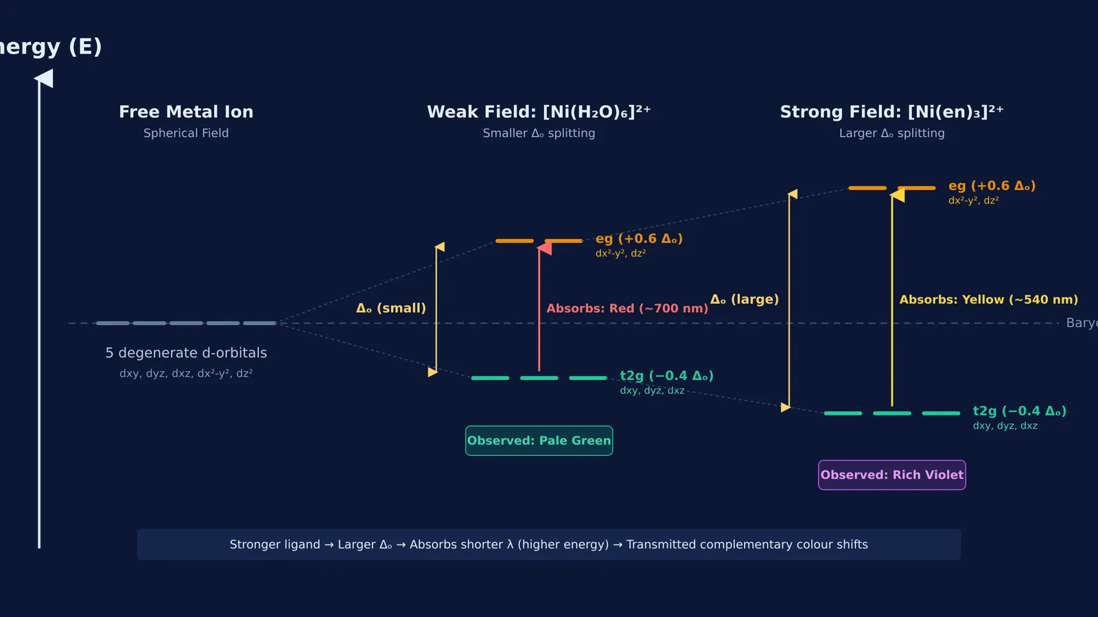
In an octahedral complex, six ligands approach along the Cartesian x, y, and z axes. Two metal orbitals—dx²-y² and dz², termed the eg set—point directly at these ligands. Direct electrostatic repulsion drives their energy up. The other three orbitals—dxy, dyz, and dxz, the t2g set—point between the axes, experience less repulsion, and drop lower.
The gap separating the two tiers is the octahedral splitting energy, Δo. To conserve total orbital energy relative to the spherical barycentre, the two eg orbitals rise by 0.6 Δo while the three t2g orbitals drop by 0.4 Δo. When an electron absorbs a photon with energy Δo, it promotes from t2g to eg. Comparing this with valence bond theory for coordination complexes shows why crystal field theory explains colour so directly.
To see how coordination geometry and ligands reshape these energy gaps in real time, explore the Fermi Simulation Lab, which provides interactive simulations for ideas that are easier to see than to read.
How do ligands shift the absorbed wavelength?
The splitting energy Δo changes with ligand identity. In 1938, chemist Ryutaro Tsuchida published absorption spectra in the Bulletin of the Chemical Society of Japan, arranging ligands into the spectrochemical series by increasing field strength:
I⁻ < Br⁻ < S²⁻ < SCN⁻ < Cl⁻ < F⁻ < OH⁻ < C2O4²⁻ < H2O < NCS⁻ < edta⁴⁻ < NH3 < en < CN⁻ < CO
Halide ions are weak-field ligands producing small splitting gaps. Chelating amines like ethylenediamine (en) and carbon donors like cyanide produce large splitting gaps.
Splitting energy determines absorbed wavelength through Planck's relation: Δo = hc/λ. Because energy is inversely proportional to wavelength, larger splitting forces the complex to absorb higher-energy, shorter-wavelength light. Using E = 1240/λ (with E in eV and λ in nm), a 2.48 eV gap absorbs cyan-blue light at 500 nm, while a 2.76 eV gap absorbs violet light at 450 nm.
How do you predict colour shifts when ligands change?
In a 2017 study in the Journal of Chemical Education, Justin M. Pratt and colleagues demonstrated that connecting particulate orbital models with synthesis helps students predict absorption shifts before measuring spectra. You can predict colour shifts using four steps:
Check d-electrons: Confirm the metal ion has an incomplete d-shell (d¹ to d⁹).
Compare ligands: Check the spectrochemical series to see if the new ligand is stronger or weaker.
Find the wavelength shift: Stronger ligands increase Δo and shorten the absorbed wavelength λ; weaker ligands decrease Δo and lengthen λ.
Read the complement: Find the colour opposite the absorbed wavelength on a colour wheel to identify the seen hue.
Consider aqueous nickel(II), a 3d⁸ cation. The hexaaqua complex [Ni(H2O)6]²⁺ has weak-field water ligands and a small Δo. It absorbs red light near 700 nm, transmitting its complement: pale green.
Adding ethylenediamine in a 3:1 molar ratio forms [Ni(en)3]²⁺. Because ethylenediamine is stronger than water, Δo widens. Absorption shifts to yellow light around 540 nm, making the solution appear rich violet. Before solving complex equilibria, review naming coordination compounds and crystal field basics.
To practise calculating splitting energies and predicting spectra across transition metal complexes, the Fermi Problem Library has worked problems with a step-by-step solver that explains the reasoning rather than printing an answer.