

On this page
In 1949, Linus Pauling and three Caltech colleagues published Sickle Cell Anemia, a Molecular Disease in Science. They showed that sickle cell haemoglobin differed from normal haemoglobin by carrying 2 to 4 more net positive charges. That was a clue, not an answer. Haemoglobin is a huge protein. Where exactly was the difference?
Seven years later, a protein chemist in Cambridge found it. His name was Vernon Ingram, and his method is still worth learning, because it shows how to find one change in a very large haystack.
Who was Vernon Ingram?
Ingram was born in 1924 in Breslau, Germany, earned a chemistry degree from Birkbeck College, London, in 1945 and a PhD in organic chemistry in 1949, according to MIT's 2006 obituary. He did the sickle cell work at the Cavendish Laboratory in Cambridge and joined the MIT faculty in 1958. MIT biology professor Graham Walker called the finding "one of the absolutely seminal discoveries in the history of molecular biology."
How did Vernon Ingram find the sickle cell mutation?
Comparing two whole haemoglobin molecules told you little beyond the charge difference. Ingram's idea was to break the problem into small pieces.
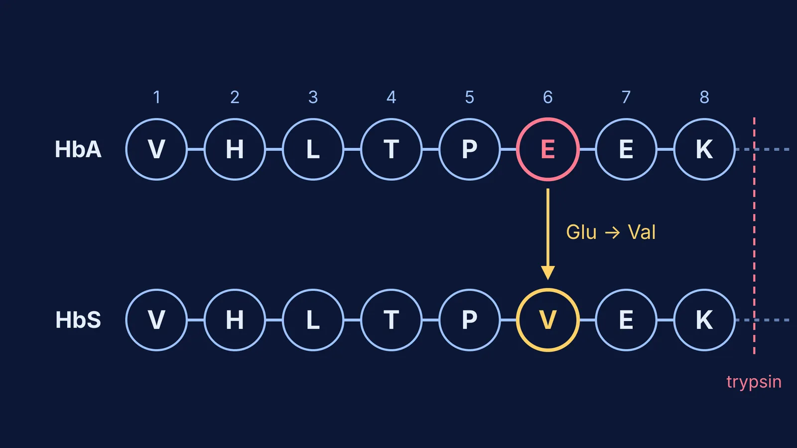
Cut. He used the enzyme trypsin to break haemoglobin at specific peptide bonds, giving a set of short peptides (University of Delaware notes on Ingram's papers).
Separate by charge. He ran the peptides along paper in an electric field (paper electrophoresis).
Separate again. He then separated them a second way, by paper chromatography.
Compare. Each peptide ended up as a spot at its own place on the paper. That pattern of spots is the "fingerprint".
The Oregon State University Pauling archive describes the result: comparing the fingerprints of normal and sickle cell haemoglobin, Ingram found that one spot differed. He reported this in Nature in 1956 in A Specific Chemical Difference Between the Globins of Normal Human and Sickle-Cell Anaemia Haemoglobin.
He then worked out the amino acids in that one peptide. In a 1957 Nature paper he showed that normal haemoglobin has glutamic acid where sickle cell haemoglobin has valine. As the Oregon State archive quotes him, the change was "a replacement of only one of nearly 300 amino-acids, namely, glutamic acid, by another valine – a very small change indeed."
Why does one amino acid change cause sickle cell disease?
Today the change is written Glu6Val: glutamic acid is replaced by valine at position 6 of beta-globin, says the US National Library of Medicine's MedlinePlus entry for the HBB gene. The same source explains that this swap makes haemoglobin S units stick together into long, rigid molecules that bend red blood cells into a sickle shape.
Glutamic acid is an acidic amino acid with a charged side chain, while valine is non-polar (our guide to classifying amino acids covers the groups). One charged side chain becomes an uncharged one, and the protein's behaviour changes.
Can you check Ingram's result against Pauling's?
Yes, and it is a good exercise in joining two papers with arithmetic.
In adults, haemoglobin has four subunits: two beta-globin and two alpha-globin (MedlinePlus).
The sickle mutation is in beta-globin, so each haemoglobin S molecule carries it twice.
Each swap removes a negatively charged glutamic acid and puts in an uncharged valine, so each beta chain becomes one unit more positive.
Two beta chains give about 2 extra net positive charges per molecule.
Pauling's group measured 2 to 4 (Delaware notes), and the Oregon State archive notes that the swap accounts for the charge difference. Two papers, seven years apart, agree.
If amino acid charges or protein subunits feel shaky, the Fermi Concept Library has a lesson for every concept in biology and chemistry, each with its place in the map, so you can fill the gap before questions like this.
How can you use Ingram's method in your own studying?
His approach works on any "what changed?" question, from a mutation problem to a wrong answer in a mock test.
Start from what is known. Ingram began with Pauling's charge difference.
Cut the big thing into comparable pieces. Peptides for him; steps of a solution for you.
Lay the two versions side by side. Normal and sickle; your working and the model answer.
Find the one spot that differs, then look closely only there.
Check the answer against the original clue. Ingram's swap explained Pauling's charge.
The Fermi Problem Library has worked problems with a step-by-step solver that explains the reasoning, which helps with step 3: put your working next to each step and find where you split off.
People who carry one copy of the variant are, according to UniProt's beta-globin entry, better protected against life-threatening malaria; we explain why in sickle cell trait and malaria. Questions about testing or health in your own family belong with a doctor.