On this page
While Rosalind Franklin is most widely known for her role in discovering the double-helix structure of DNA, her subsequent research on viruses was just as ground-breaking. Moving to Birkbeck College in 1953, she turned her X-ray crystallography expertise to the tobacco mosaic virus (TMV).
Her meticulous experimental work shifted how the scientific world saw viruses. Rather than mysterious, formless infectious agents, Franklin showed them to be precise, orderly molecular machines built from repeating parts.
What is the tobacco mosaic virus structure?
Before Franklin's measurements, scientists knew TMV was rod-shaped, but its internal architecture was a mystery. Some thought the virus might be a solid cylinder of protein, while others guessed the genetic material acted like a wick running down the exact centre.
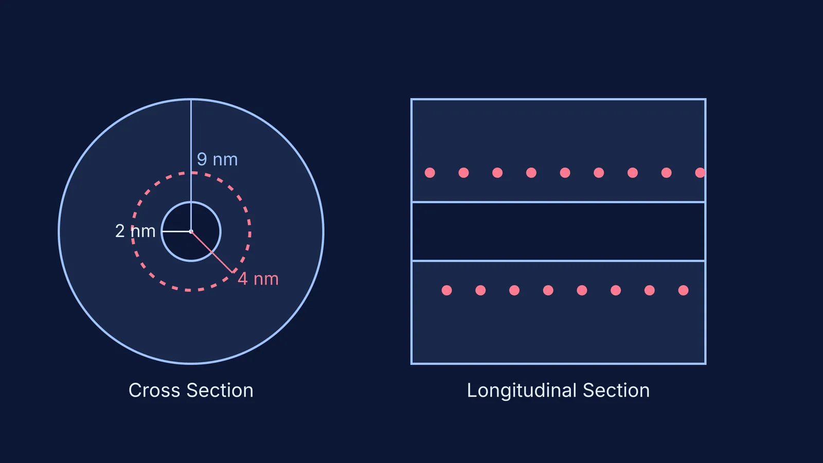
Franklin established that TMV is a rigid, hollow cylinder, approximately 300 nm long with an outer diameter of 18 nm. Most surprisingly, she found a central, water-filled channel running down the axis with a diameter of 4 nm.
The protective outer shell, known as the capsid, is built from roughly 2,130 identical protein subunits. These subunits do not form independent, stacked rings; instead, they lock together in a continuous right-handed helix, winding upwards with a pitch of 2.3 nm per turn.
Where is the RNA located in the virus?
In a landmark 1956 paper, Franklin proved that the virus's genetic material—a single strand of RNA—was neither at the very centre nor wrapped around the outside of the particle.
Instead, the RNA is deeply embedded within the protein subunits, winding in a corresponding helix at a precise radius of 4 nm from the central axis.
By burying the RNA inside the thick protein wall, the virus ensures its genome survives until it enters a susceptible host. Unlike RNA processing in eukaryotes, which happens safely inside a cell nucleus, a viral genome must survive the harsh extracellular environment. If you are trying to picture complex molecules or helical structures, the Fermi Simulation Lab has interactive simulations for the ideas that are easier to see than to read.
How did Rosalind Franklin map the virus?
Franklin mapped TMV using high-resolution X-ray crystallography. By shining an intense beam of X-rays through highly ordered, crystallised virus samples and studying the diffraction patterns they cast on photographic film, she could work backwards to calculate the distances between atoms.
This process was exceptionally demanding. Before modern computers, the mathematical transformations required to turn spots on a film into a three-dimensional model had to be calculated by hand. Because TMV is highly symmetrical, the diffraction patterns it produced were exceptionally clear. However, interpreting these complex patterns required extraordinary mathematical skill, a deep understanding of physical chemistry, and immense spatial reasoning to visualise the molecule in three dimensions.
When studying biology, building a strong mental model of what you are reading is essential, whether you are trying to classify viruses, viroids and lichens or visualising microscopic structures. Fermi's AI tutor can work out what you already know and build a plan for the session, so you sit down to a clear next step instead of feeling overwhelmed by complex spatial arrangements.
What was Franklin's legacy beyond DNA?
Franklin continued to publish on the tobacco mosaic virus and began pioneering work on the polio virus before her untimely death in 1958.
Her structural models were so definitive that she was invited to build a large-scale physical model of TMV for the 1958 Brussels World's Fair. Furthermore, her meticulous X-ray diffraction data laid the foundation for her colleague Aaron Klug. He continued her structural biology work to fully map the virus, research for which he later received the Nobel Prize in Chemistry in 1982.
Ultimately, Franklin's research transformed virology. She proved that the rigorous tools of physical chemistry could unravel the invisible architecture of life, revealing the elegant geometric principles that govern how viruses assemble and function.