On this page
In 1962, Japanese chemist Osamu Shimomura was trying to understand why a small jellyfish glowed in the dark 1. He did not know that he was about to give biology its most useful light bulb.
Shimomura isolated green fluorescent protein (GFP) from the jellyfish *Aequorea victoria* 1. Today, this protein is attached to other molecules in laboratories worldwide, acting as a molecular lantern that lets scientists watch cancer cells spread, nerves develop, and viruses multiply in real time.
What did Osamu Shimomura discover?
Shimomura arrived at Princeton University in 1960 and soon began studying bioluminescence with his colleague Frank Johnson 2. Shimomura had already made a name for himself in Japan; in 1955, working at Nagoya University, he successfully crystallized the light-emitting molecule from a small crustacean called *Cypridina* 2.
At Princeton, the focus shifted to *Aequorea victoria*, a jellyfish that lights up with a green ring around its edge. To collect them, the team travelled to Friday Harbor Laboratories in Washington.
Their first discovery in 1962 was not green at all 1. They isolated a protein that emitted a faint blue light when calcium ions were added to it, which they named aequorin.
During the purification process of aequorin, Shimomura noticed another trace protein that did not glow on its own but emitted a bright green light when exposed to ultraviolet light. He had discovered green fluorescent protein. The sheer scale of the effort was staggering. Between 1961 and 1988, Shimomura and his team—including his wife Akemi—caught approximately 850,000 jellyfish, processing up to 3,000 a day in a dedicated effort to extract enough of these proteins to deduce their chemical structure 2. By 1979, he had finally elucidated the chemical structure of the part of the GFP molecule that glows, known as the chromophore 2.
How does the jellyfish Aequorea victoria glow?
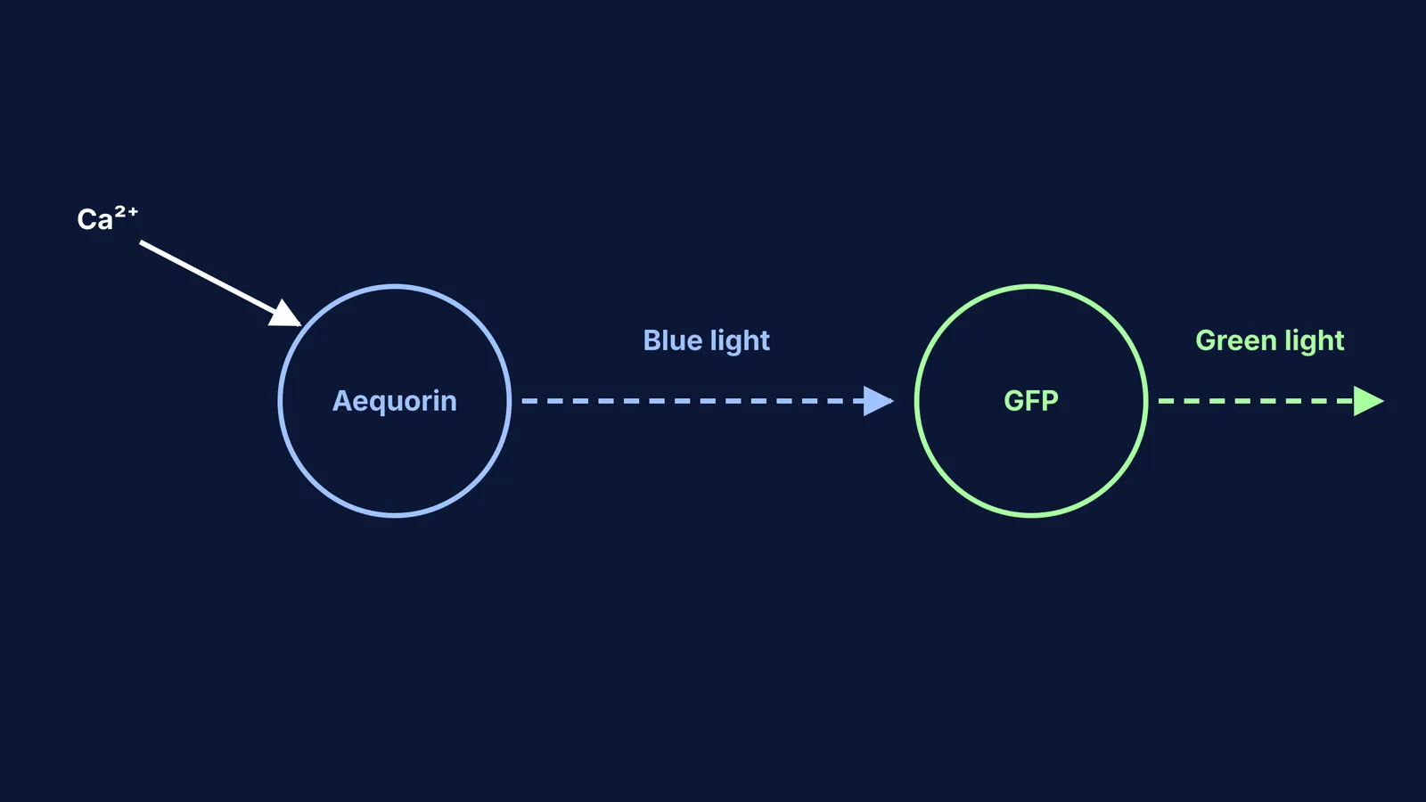
The jellyfish creates its green glow through a two-step process 1. First, calcium ions enter the cell and bind to aequorin, causing it to release blue light.
Then, the nearby GFP absorbs this blue light and re-emits it as a lower-energy green light. This transfer of energy explains why the jellyfish looks green in the water, even though its primary light-producing chemical emits blue.
If you are curious about how complex molecules like aequorin and GFP form, Protein structure from primary to quaternary, with worked numbers explains the stages of folding.
Why is green fluorescent protein useful?
For decades, GFP was just a biological curiosity. Then, in the 1990s, researchers realised they could stitch the gene for GFP onto the genes for other proteins 1.
Because GFP forms its glowing core spontaneously without needing extra enzymes or chemical cofactors, it acts as a permanent, built-in torch. When the cell reads the DNA and produces the target protein, it produces the attached GFP too. Scientists can shine a light on the cell and watch exactly where the protein goes, how fast it moves, and what it interacts with.
This technique lets researchers track almost anything inside a living organism. It has been used to follow the spread of cancer cells, watch how HIV replicates, map the development of nerve cells in the brain, and detect environmental pollutants like cadmium, zinc and even explosive fumes 1. Before this, tracking proteins often meant killing the cell and using radioactive tracers or complex chemical dyes that could interfere with the organism's natural processes. With GFP, biology became a real-time movie rather than a static photograph.
Other scientists, building on the work of pioneers like Margaret Dayhoff and the first protein sequence database, later mutated GFP to glow in cyan, yellow and red. This allowed laboratories to track multiple different proteins at the same time, turning cells into colourful, observable machines.
A framework for understanding fluorescent tagging
When reading about biological markers or tracking proteins in your biology syllabus, you can use the TAG framework to understand the experiment:
Target: What is the protein or gene the scientist wants to study?
Anchor: How is the marker (like GFP) attached? Usually, the DNA sequence for GFP is fused to the DNA sequence of the target protein.
Glow: What triggers the marker to light up? For GFP, it only requires shining a specific wavelength of light onto the sample.
This simple breakdown helps decode complex molecular biology questions in exams like NEET or school practicals. If you need to revise the basics of how these molecules interact, the Fermi Concept Library has every concept in physics, chemistry, biology, mathematics and computing, each with its own lesson and its place in the map.
If you are studying molecular biology and find yourself struggling to connect these ideas, Fermi's AI tutor can work out what you already know and build a plan for the session, so you sit down to a clear next step.