On this page
When the First World War began in 1914, X-rays had been known only since 1895. In her book La Radiologie et la Guerre (1921), Marie Curie says France's radiology services were notoriously inadequate, and many emergency hospitals in schools had no electricity at all. Her answer was to bring the electricity, the machine and trained people to the wounded.
What were Marie Curie's X-ray cars?
In her book, Curie explains that she ran the X-ray work of two relief bodies, the Patronage National des Blessés and the Red Cross's Union des Femmes de France. The first car, from the Union, served Paris during the rush of wounded after the Battle of the Marne in September 1914. By her own count she equipped 18 X-ray cars with private donations and Patronage funds, and the Patronage set up about 200 fixed X-ray posts in hospitals.
Timothy Jorgensen of Georgetown University writes in The Conversation that Curie soon had 20 cars, that they were nicknamed little Curies, that she learned to drive and change flat tyres, and that an estimated one million-plus wounded soldiers had X-ray exams during the war.
How did a mobile X-ray unit work?
Curie describes the cars in detail in La Radiologie et la Guerre:
Power: the car's engine could drive a dynamo on the running board. A separate generator weighed at least 100 kg; the dynamo halved the weight.
Crew: a doctor, an X-ray operator and a driver.
Set-up: a 25-metre cable into the ward, blankets over the windows, often ready in half an hour.
Range: early on, a radius of more than 100 km, at up to 50 km per hour.
The X-rays answered what surgeons could not see. Was a bullet still inside, and exactly where? How was a broken bone lying? Curie writes that fractures took most of the time hospitals spent on the wounded, and that without X-rays surgeons often could not attempt to remove a bullet.
Who did Curie train to run the X-ray units?
In her book, Curie says she started a school for women X-ray operators at the Radium Institute in Paris, which trained about 150 women from 1917 to the end of the war. Each course took about 20 students for six weeks to two months, all day. Theory covered electricity, current and voltage, induction and the X-ray tube; practice meant running real equipment, plus basic anatomy and reading X-ray plates. Jorgensen adds that her daughter Irène taught with her.
How did X-rays find the depth of a bullet?
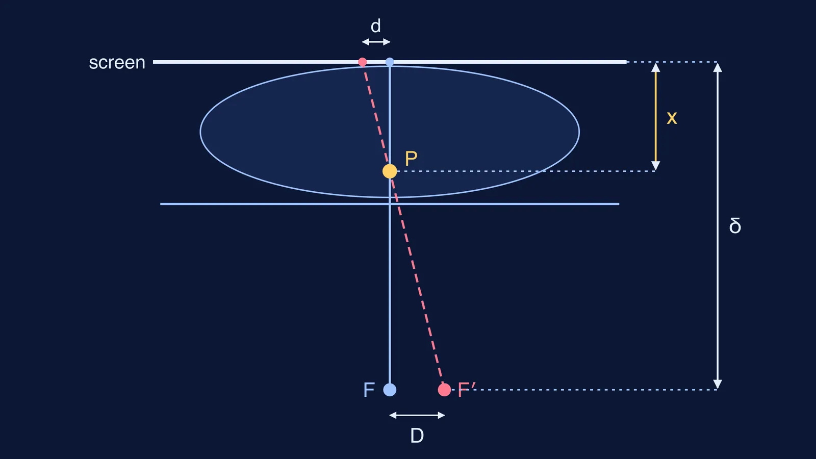
A single X-ray is a shadow: it shows where a bullet sits, but not how deep. Curie's double-image method uses two shadows. Centre the tube under the bullet, mark the skin, slide the tube a known distance D (10 cm, for example), and measure how far the shadow moves on the screen, d. If δ is the distance from the tube's focus to the screen, the bullet's depth below the screen is x = δ × d ÷ (d + D).
Worked example (numbers chosen for practice):
The tube focus is 60 cm below the screen, so δ = 60 cm.
You slide the tube D = 10 cm and the shadow moves d = 5 cm.
x = 60 × 5 ÷ (5 + 10) = 20 cm below the screen.
Subtract the small gap between screen and skin to get the depth below your skin mark.
Why it works: the bullet sits δ − x above the tube, and the triangle of two tube positions and bullet is similar to the triangle of two shadows and bullet, so d ÷ D = x ÷ (δ − x). Rearrange to get Curie's formula. It holds only while the patient keeps still.
If similar triangles still feel slippery, drawing both rays helps. The Fermi Simulation Lab has interactive simulations for ideas that are easier to see than to read, and the Fermi Problem Library has worked problems with a step-by-step solver that explains the reasoning.
How can you try Curie's method at home?
Test the same geometry with a torch and a pencil:
In a dark room, stand a pencil 20 to 30 cm from a wall and shine a small torch at it from about 1 m from the wall.
Mark the pencil's shadow on paper taped to the wall.
Slide the torch 10 cm sideways and mark the new shadow.
Measure the shift d and use x = δ × d ÷ (d + D), with δ as the torch-to-wall distance, to predict the pencil's distance from the wall.
Check with a ruler. If you are off, ask which measurement was least precise.
Why a torch and not real X-rays? Jorgensen writes that Curie worried her intense X-ray work would harm her, and years later she developed aplastic anaemia, a blood disorder sometimes caused by high radiation exposure. Light and shadows teach the same geometry safely.
Read Curie's own account, with her Fig. 10, free on Project Gutenberg.