

On this page
In June 2014, Manu Prakash's lab at Stanford described a microscope you fold from paper. Their paper, Foldscope: Origami-Based Paper Microscope in PLOS ONE, by James Cybulski, James Clements and Prakash, reports that it costs less than a dollar in parts and can give over 2,000× magnification.
Prakash's MacArthur Foundation fellow profile lists a B.Tech from IIT Kanpur in 2002 and a PhD from MIT in 2008, and he was named a MacArthur Fellow in 2016.
What is the Foldscope?
The Foldscope is an optical microscope folded from a single sheet of paper, with the lenses and electronics built in, as the MacArthur profile describes it. The 2014 paper gives the specifications:
It weighs 8.8 grams and measures 70 × 20 × 2 mm.
It can be folded together from a flat sheet in under 10 minutes.
Its parts include a ball lens, an LED, a button-cell battery, a switch, copper tape and paper stages.
The LED uses only 6 mW and can run for over 50 hours on a CR2032 coin cell.
It can survive being dropped from a three-storey building or stepped on.
How does the Foldscope paper microscope work?
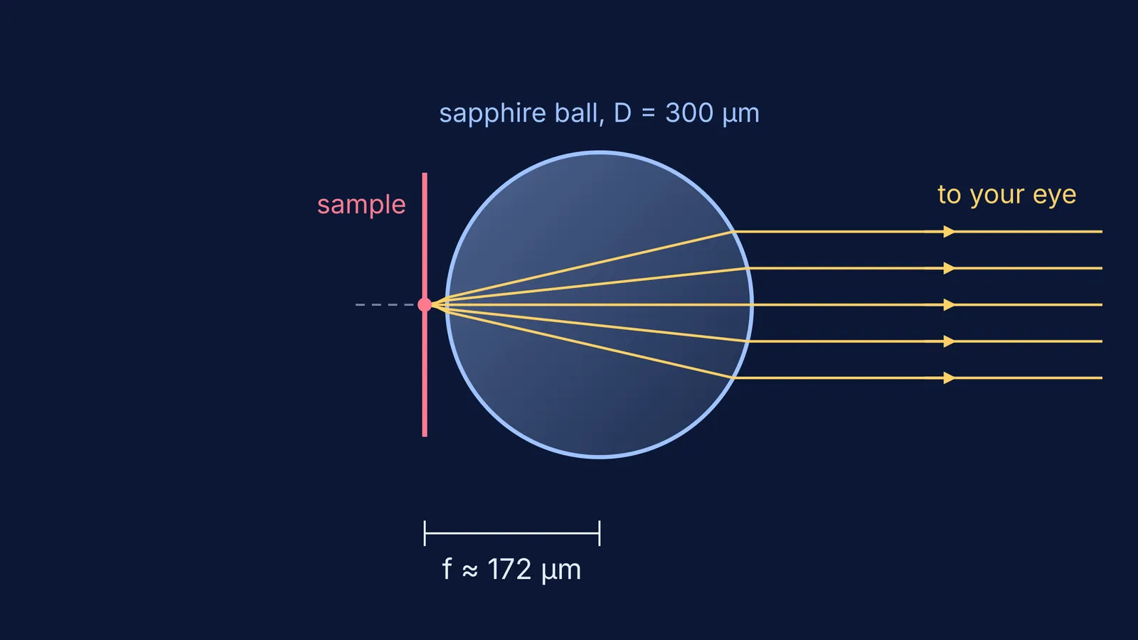
Everything is built around one tiny sphere of glass, sapphire or ruby (PLOS ONE).
The lens. The ball sits in a hole in the paper. One bead on its own is a complete microscope, the same idea as the single-lens instruments Antonie van Leeuwenhoek used to see bacteria.
The sample. You mount your specimen on a paper slide (it can be a wet mount for live organisms) and slide it into the frame.
Focusing. Pulling your thumbs apart or pushing them together flexes the paper stage, which moves the lens towards or away from the sample. The paper calls this flexure-based focusing.
Panning. Moving both thumbs together slides the lens and the light across the slide. The sample stays fixed while the optics move.
Viewing. You hold the Foldscope in both hands and, in the paper's words, place your eye close enough to the lens "so one's eyebrow is touching the paper".
Why does a tiny bead magnify so much?
A smaller sphere has a shorter focal length, which means more magnification. A ball lens of diameter D and refractive index n has an effective focal length EFL = nD / 4(n − 1), measured from its centre (ball lens formula). A simple magnifier is rated as M = 250 mm / f, because 25 cm is the standard viewing distance (magnifier formula).
Here is the worked example for the Foldscope's 300 µm sapphire lens, using n ≈ 1.77, the value the paper uses:
EFL = (1.77 × 300 µm) / (4 × 0.77) ≈ 172 µm.
Back focal length = EFL − D/2 = 172 − 150 = 22 µm. The sample sits just 22 µm from the bead's surface.
Magnification = 250 mm / 0.172 mm ≈ 1,450×.
These match the paper's figures for this lens (EFL 172 µm, BFL 22 µm, 1,450X), which also reaches submicron resolution.
Your turn: a 1 mm glass bead with n = 1.5 gives EFL = 1.5 × 1 / (4 × 0.5) = 0.75 mm, so M = 250 / 0.75 ≈ 330×. Our worked guide to the magnifying power of microscopes and telescopes has board-exam versions of these problems.
What can you see with a Foldscope?
The 2014 team imaged parasites including Giardia lamblia, Leishmania donovani and Trypanosoma cruzi, and the bacteria Escherichia coli and Bacillus cereus, at magnifications from 140X to 2,180X (PLOS ONE). On a standard resolution target, their 430X and 140X lenses resolved 1.38 µm and 2.19 µm.
The Foldscope came to India after the Department of Biotechnology (DBT) signed a Statement of Intent with Prakash Lab on 27 September 2015. The DBT Foldscope programme page, hosted by DBT-promoted Biotech Consortium India Limited, reports that DBT has supported more than 400 schools and colleges, and that these ran 2,020 workshops and trained 117,573 students.
How do you get the most from a pocket microscope?
Start with the weakest lens, find the sample, then switch to a stronger one.
Use a thin sample. With a depth of field of a few microns (PLOS ONE), a thick slice just looks blurred.
Sketch what you see and label it. Drawing makes you notice structure, as Robert Hooke did when he named the cell.
Work out the scale before you claim a size. With the 300 µm lens, the sharpest part of the view is only about 176 µm across (an 88 µm radius), so anything that fills it is under a fifth of a millimetre.
If refraction through a sphere is hard to picture, the Fermi Simulation Lab has interactive simulations for ideas that are easier to see than to read. For lens numericals, the Fermi Problem Library has a step-by-step solver that explains the reasoning rather than printing an answer.


