On this page
Reading DNA used to mean a scientist looking at dark bands on X-ray film and writing the letters down by hand. In June 1986 a Caltech team led by Leroy Hood showed how to hand that job to a laser and a computer. The trick was to give each of DNA's four bases its own colour of fluorescent dye.
What did Leroy Hood's automated DNA sequencer do?
The method appeared in the journal Nature as Smith, Hood and colleagues, "Fluorescence detection in automated DNA sequence analysis" (1986). Lloyd Smith was the first author and Leroy Hood the senior author. Their abstract calls it a method for the partial automation of DNA sequence analysis.
It built on the enzymatic method of Frederick Sanger. In that method, copies of a DNA strand stop growing at a particular base, which gives a set of fragments of different lengths. Older versions labelled these fragments with radioactivity, and the result was read from an autoradiogram. The NHGRI Human Genome Project fact sheet shows Francis Collins studying one.
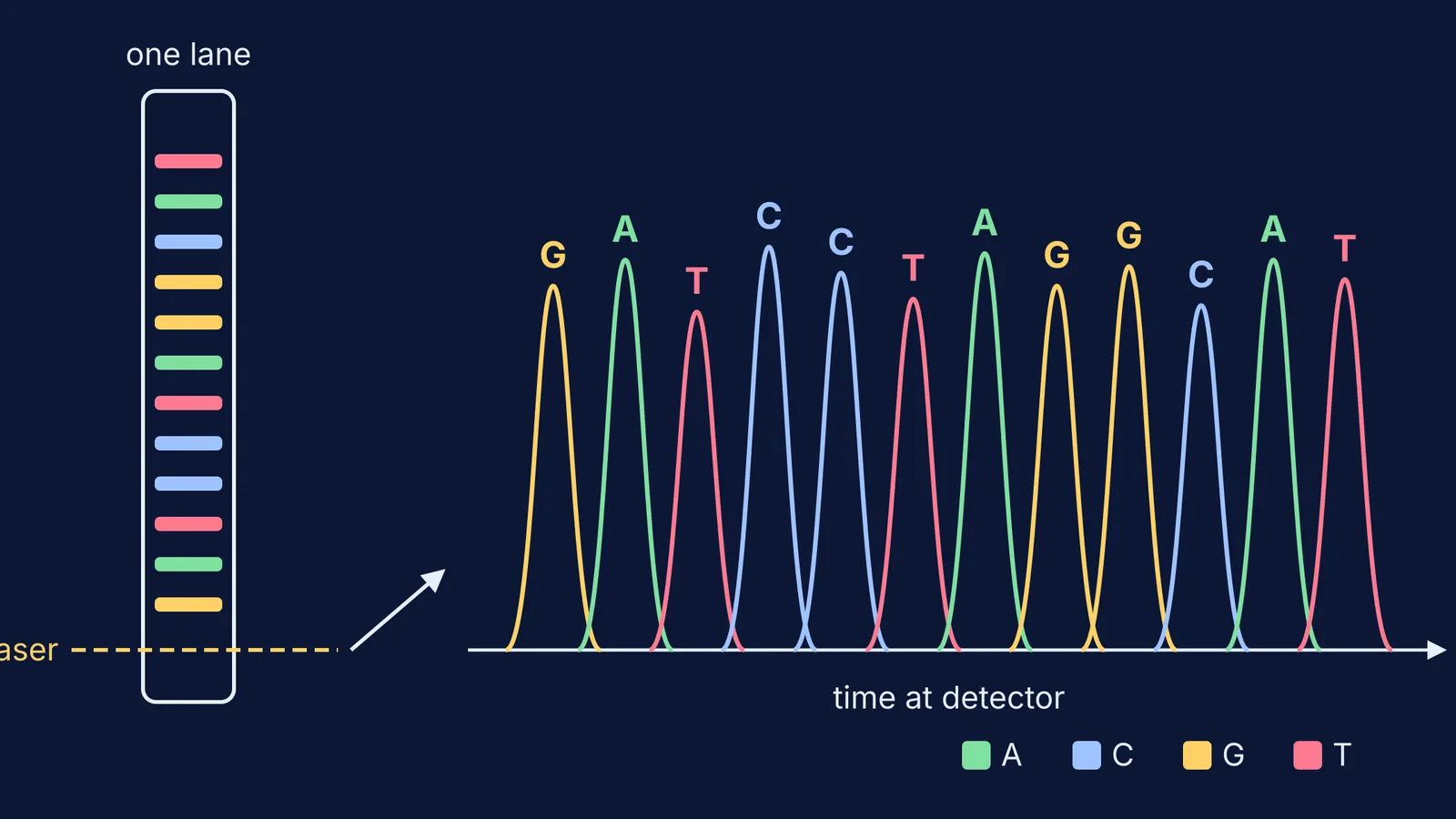
According to the 1986 abstract, the Caltech team attached a fluorescent dye to the primer, the short piece of DNA where copying begins. They used a different colour in each of the reactions for A, C, G and T. All four reaction mixtures were then combined and run down one polyacrylamide gel tube. A detector near the bottom saw each coloured band go past, and the sequence went straight into a computer.
How did four fluorescent dyes speed up sequencing?
Colour did the sorting. If each base has its own colour, the machine only needs to record which colour arrives next. The Lemelson-MIT Program describes the process as a laser making the DNA glow red, green, blue or orange, with a computer reading the signals.
Run four copying reactions, one per base, each with a primer carrying its own dye.
Mix them and run them together through a single gel. Shorter fragments travel faster.
Shine a laser across the gel near the bottom so each band glows as it passes.
Let the computer log the colour sequence: that is the DNA sequence.
The Lemelson-MIT Program says Hood's team built a prototype at Caltech in 1985. A prototype instrument made by Applied Biosystems in 1987 is now in the Science Museum Group collection in the UK. The museum describes it as using the Sanger method, with the sequence read by a laser and shown on a screen or printout.
How did the sequencer help the Human Genome Project?
According to the NHGRI Human Genome Project fact sheet, the project ran from 1990 to 2003 and its initially projected cost was $3 billion. NHGRI says the project relied on Sanger sequencing, greatly advanced through a series of technical innovations.
Hood and Lee Rowen, writing in Genome Medicine in 2013, describe step-by-step improvements in the cost, throughput, speed and accuracy of automated fluorescent sequencing throughout the project. They note that the initial human genome reference sequence was read on a 96-capillary version of this first-generation technology. The Lemelson-MIT Program puts it more simply: over 17 years the machine made DNA sequencing 3,000 times faster. For the next big step, see how Craig Venter used shotgun sequencing on the first bacterial genome.
How do you read a four-colour sequencing trace?
Here is a worked example. Suppose the trace, from the shortest fragment to the longest, shows peaks in this colour order, with A green, C blue, G yellow and T red:
yellow, green, red, blue, blue, red, green, yellow, yellow, blue, green, red
Turn colours into letters: G A T C C T A G G C A T.
This is the new strand, read 5' to 3', because copying adds bases to the 3' end and the shortest fragment arrives first.
Pair each base (A with T, G with C) to get the template, which runs the other way: 3' C T A G G A T C C G T A 5'.
Flip it so it reads 5' to 3': ATGCCTAGGATC.
If it is hard to picture fragments racing through a gel, the Fermi Simulation Lab has interactive simulations for ideas that are easier to see than to read. It also helps to revisit how gel electrophoresis separates DNA fragments.
What can students learn from Hood's sequencer?
The 1986 paper joined chemistry (dyes), physics (lasers and fluorescence) and computing so that a machine, not a person, read the bands. Use this quick check before a biology test:
Can you explain why shorter fragments reach the detector first?
Can you say what the dye in the 1986 method was attached to?
Can you turn a colour trace into the template strand, with the 5' and 3' ends labelled?
If any of these feels shaky, the Fermi Concept Library covers every concept in biology and chemistry, each with its own lesson. Try the worked example again tomorrow without looking at the answer.