

On this page
Before the 1980s, the only way to see the three-dimensional shape of a protein was through X-ray crystallography. This technique, used by scientists like Margaret Dayhoff and Kathleen Lonsdale, required coaxing the protein into a solid, highly ordered crystal. While powerful, crystals force proteins into a rigid state that does not perfectly match their natural environment. Inside a living cell, proteins float, fold and interact in a watery solution.
Kurt Wüthrich, a Swiss chemist, developed a way to see proteins in this natural liquid state. For his work in developing nuclear magnetic resonance (NMR) spectroscopy for determining the 3D structure of biological macromolecules in solution, he was awarded half of the 2002 Nobel Prize in Chemistry (NobelPrize.org).
Why study proteins in solution?
Proteins are not static shapes; they are dynamic molecules that flex and move to do their jobs. When forced into a crystal, some of this flexibility is lost, and the true shape may be slightly altered by the tight packing of the crystal lattice. Some proteins, such as prions, are also notoriously difficult to crystallise.
By studying proteins in an aqueous solution, researchers observe them under conditions closer to those inside a human body. This provides a more accurate picture of how they fold, how they interact with other molecules, and how drugs might bind to them to treat diseases.
How does NMR find protein structure?
Nuclear magnetic resonance (NMR) relies on the fact that certain atomic nuclei, such as the protons found in hydrogen atoms, act like tiny spinning magnets. When a sample is placed in a very strong magnetic field and hit with pulses of radio waves, these nuclei absorb and re-emit energy at specific frequencies. The exact resonance frequency, or "chemical shift", depends on the local electron cloud surrounding the atom. Because a protein contains hundreds or thousands of hydrogen atoms, its one-dimensional NMR spectrum is initially just a crowded, overlapping forest of peaks.
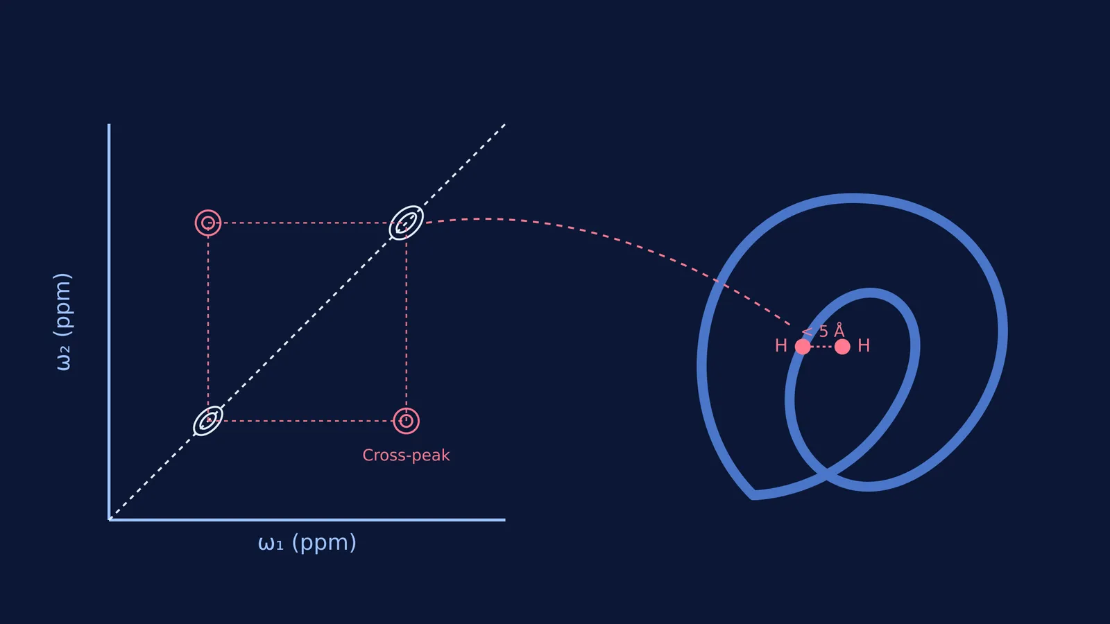
Wüthrich's breakthrough in the early 1980s was developing a systematic way to assign these radio signals to specific atoms in a large protein chain. He used a phenomenon called the Nuclear Overhauser Effect (NOE). When two protons are very close to each other in space—typically less than 5 Ångströms (5 Å) apart—their magnetic properties interact. This interaction shows up as a "cross-peak" in a two-dimensional NMR spectrum (Journal of Biomolecular NMR).
Even if two hydrogen atoms are far apart in the linear sequence of the protein—perhaps one is at the beginning of the chain and the other is near the end—they might be brought close together when the protein folds into its complex 3D shape. By systematically measuring thousands of these short-distance interactions, Wüthrich and his team created a vast web of distance constraints. Because the covalent bonds linking the atoms are already known, a computer algorithm can use this geometric puzzle to calculate the only possible 3D structures that satisfy all the distance rules. Instead of a single static model, NMR often produces an "ensemble" or a family of closely related structures, reflecting the natural wiggling and breathing of the protein in the liquid.
The shape of the prion protein
One of the most famous applications of Wüthrich's method was determining the structure of the prion protein. Prions are the proteins responsible for transmissible spongiform encephalopathies, such as mad cow disease and its human equivalent, Creutzfeldt-Jakob disease. The infectious form of a prion is a misfolded version of a normal cellular protein.
Because prions are notoriously difficult to study with crystallography due to their flexibility, Wüthrich's team at ETH Zürich used NMR to solve the structure of the healthy cellular prion protein in 1996 (Nature). They discovered that the protein has a unique two-part architecture. The front half of the chain is a long, flexibly disordered tail that flops around freely in solution. The back half is a well-defined globular domain containing three coiled alpha-helices and a short, two-stranded beta-sheet.
By revealing the healthy structure, NMR provided the essential baseline for researchers trying to understand how the protein flips into its deadly, disease-causing shape. This work proved that NMR could not only handle standard proteins, but could also map highly flexible molecules that lack a single static form.
The impact of solution NMR
Following his breakthrough in the early 1980s, Wüthrich's method proved that NMR could be a viable alternative to crystallography for determining atomic-resolution structures. Since then, thousands of protein structures have been solved using NMR and added to global repositories.
Today, X-ray crystallography and NMR spectroscopy are complementary tools. Crystallography handles large, complex assemblies, while NMR excels at mapping smaller, highly dynamic proteins in their natural liquid environment. If you are learning how physical techniques reveal molecular shapes, the Fermi Concept Library breaks down the physics of magnetic resonance, and the Fermi Simulation Lab lets you interact with 3D protein folds.
How to read a 2D NMR spectrum
If you encounter a 2D NOESY (Nuclear Overhauser Effect Spectroscopy) plot in a chemistry or biochemistry exam:
Find the diagonal: The strong signals running from the bottom-left to the top-right represent the standard chemical shifts of the protons.
Look for off-diagonal peaks: These are the cross-peaks. They appear symmetrically on either side of the diagonal.
Trace them back: A cross-peak at coordinates (x, y) means that the proton with chemical shift x is close in space to the proton with chemical shift y.
Use the intensity: A darker or more intense cross-peak indicates a shorter distance between the two protons.


