

On this page
In 1839, the Czech physiologist Jan Evangelista Purkyně was examining the inner walls of the heart when he noticed a network of distinct, greyish threads just beneath the surface. Today, we know these as the Purkinje fibres—the terminal branches of the heart's electrical conduction system, responsible for keeping our heartbeat synchronized and powerful.
Purkyně was one of the 19th century’s greatest microscopists. By the time he found the heart fibres, he had already identified the large Purkinje cells in the cerebellum of the brain in 1837 (Wikipedia).
What did Jan Purkyně see in the heart?
Using the microscopes of the 1830s, Purkyně saw that these subendocardial fibres were morphologically distinct from regular contractile heart muscle. At the time of his 1839 discovery, the concept of electrical conduction in the heart was largely unknown, and he initially suspected these structures might be related to the nervous system (Wikipedia).
Today, we know these fibres appear lighter and larger than their neighbors on a microscope slide because of the presence of glycogen around the nucleus (Wikipedia). It took decades for later scientists to realise they were not structural supports or nerves, but rather the heart's high-speed electrical wiring.
How do Purkinje fibres make the heart beat?
A heartbeat is fundamentally a wave of electricity followed immediately by a wave of muscle contraction. The signal starts at the top of the heart in the sinoatrial (SA) node, which acts as the natural pacemaker. It then spreads down and pauses briefly at the atrioventricular node discovered by Sunao Tawara. This tiny delay allows the atria to fully empty their blood into the ventricles below.
From the AV node, the signal races down the centre of the heart through the bundle of His and splits into the left and right bundle branches. When it reaches the very bottom of the ventricles (the apex), it transfers into the extensive network of Purkinje fibres.
These fibres are built explicitly for speed. Purkinje fibres conduct cardiac action potentials more quickly and efficiently than any other cells in the heart's electrical conduction system (Wikipedia). Because they act as a motorway for the electrical signal, they deliver it to the thick ventricular muscles almost instantaneously.
Mapping the cardiac cycle in your head is hard. The Fermi Simulation Lab has interactive simulations for ideas that are easier to see than read, letting you build visual models of complex invisible processes.
What makes the Purkinje action potential unique?
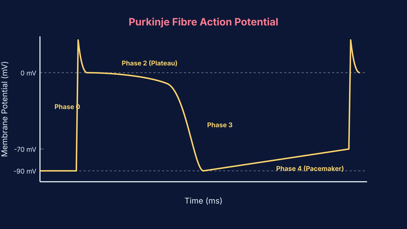
Unlike regular skeletal muscle, cardiac cells have distinct electrical signatures known as action potentials (Wikipedia). The action potential of a Purkinje fibre is unique because of its extended duration and specific sequence of ion movements.
When the electrical signal arrives, voltage-gated sodium channels burst open, causing a sharp, nearly vertical spike in membrane potential (Phase 0). This is followed immediately by a small notch (Phase 1) as some potassium exits the cell. The most critical part of the cycle is Phase 2, the plateau. During this phase, calcium channels open, allowing calcium ions to slowly enter the cell. This balances the exiting potassium and keeps the cell depolarized for hundreds of milliseconds. This long plateau prevents the heart from going into tetanus—a sustained, locked contraction—which would be fatal.
After the plateau, the calcium channels close, and the cell rapidly returns to its resting state (Phase 3). But in Purkinje fibres, the resting state is never truly at rest. During Phase 4, a slow, continuous leak of sodium and calcium ions gradually brings the membrane potential back up. This pacemaker potential is what allows the fibres to trigger their own beat if they do not receive a signal from the AV node.
Why must the ventricles contract from the bottom up?
The Purkinje fibres branch out from the apex and sweep upward along the outer walls of the ventricles. Because the electrical signal reaches the bottom of the heart first, the muscle contraction starts at the apex and moves upward toward the base.
This specific sequence is a mechanical necessity. The heart's exit valves—the aortic valve and the pulmonary valve—are located at the top of the ventricles. By squeezing from the bottom up, the heart efficiently forces blood out of the right ventricle for pulmonary circulation first described by Ibn al-Nafis, and out of the left ventricle to the rest of the body.
If the ventricles contracted from the top down, or even all at once, blood would simply be trapped against the closed bottom of the chamber. The apex-to-base sequence ensures that maximum pressure is directed exactly where the blood needs to go.
What happens if the main pacemaker fails?
The Purkinje network also serves as the heart's ultimate backup plan. If the SA node and the AV node both fail to send a signal, the Purkinje fibres possess their own automaticity. They will spontaneously generate an electrical impulse at an intrinsic rate of 20 to 40 beats per minute (Wikipedia). While this rate is too slow to support intense physical activity, it provides a crucial safety net that can keep a person alive until medical help is administered.


