On this page
From 1929 to 1970, Honor Fell directed the Strangeways Research Laboratory in Cambridge. There, she perfected the "organ culture" technique. While traditional cell cultures grew dissociated cells in flat, two-dimensional sheets that lost their original form and function, Fell wanted to see how tissues behaved in their natural three-dimensional shapes.
By placing explants like embryonic limb buds on a solid clot of plasma and embryo extract in a watch-glass, she kept the tissue's architecture intact. This semi-solid surface provided the physical support the cells needed to maintain their spatial relationships. This allowed her to watch a tiny, unformed cartilage rudiment slowly grow and ossify into a recognisable bone, entirely outside a living body, much like the ones you would count when studying The human skeleton: counting axial and appendicular bones. Her work proved that the pattern for skeletal development is intrinsically programmed into the tissue itself, rather than being shaped entirely by external forces in the body.
What does vitamin A do to developing cartilage?
In their 1952 study, Fell collaborated with Sir Edward Mellanby, a nutritionist who had previously studied vitamin deficiencies (see Edward Mellanby and the puppy diets that explained rickets). They set out to observe the direct effects of hypervitaminosis A (excess vitamin A) on tissues.
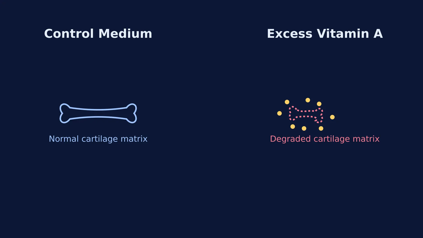
They added high concentrations of vitamin A to the culture medium of embryonic chick limb bones. The result was striking: the cartilage matrix rapidly broke down and resorbed, causing the bones to become soft and lose their structural integrity. The bone literally appeared to melt away over a few days. Fell suggested that the cells were releasing powerful destructive enzymes to break down their own matrix in response to the vitamin. This observation laid the groundwork for understanding lysosomes—the cell's recycling centres that store these enzymes—and their role in tissue degradation. This discovery added another vital layer to our understanding of cell organelles alongside the work of George Palade and the ribosomes in the cell's protein factory.
If you are trying to understand the sequence of endochondral ossification (how cartilage turns into bone) and where these enzymes act, the Fermi Concept Library covers every concept in biology to help you trace the development step by step.
Can a cell change its fate completely?
In a 1953 experiment, Fell and Mellanby tested vitamin A on 6- to 7-day-old chick embryo skin (ectoderm). Normally, this embryonic tissue develops into a tough, dry, keratinized layer, forming structures like scales or feathers that protect the outside of the animal.
However, when grown in a medium with high vitamin A, the tissue underwent a dramatic transformation called mucous metaplasia. Instead of producing rigid keratin, the skin cells transformed into a ciliated, mucus-secreting epithelium, physically identical to the lining of the respiratory tract. The cells even developed tiny beating hairs (cilia) that moved the secreted mucus across the tissue surface.
This was a landmark discovery in developmental biology. It proved that a single chemical factor could completely switch the developmental pathway of a tissue in a controlled environment. The finding demonstrated that the fate of embryonic cells is not rigidly fixed but responds dynamically to chemical signals, challenging the idea that cells were locked into their developmental paths.
How do you learn developmental biology pathways?
Developmental biology relies on understanding how chemical signals change physical structures over time. When studying these experiments, use a structured approach to keep track of the changes rather than trying to memorise isolated facts:
Trace the signal: Identify the specific hormone or vitamin applied to the tissue (e.g., vitamin A).
Observe the structural shift: Note how the three-dimensional arrangement or cell type changes (e.g., cartilage breaking down, or keratinized skin turning into mucus-secreting tissue).
Identify the mechanism: Look for the cellular machinery responsible for the change (e.g., the release of proteases from lysosomes dissolving the matrix).
When you are working through complex biological processes, it helps to have a system that holds a line of reasoning across your entire study session. Fermi's AI tutor can work out what you already know about a pathway and build a plan for the session, checking your understanding of the mechanisms before moving on to the final structural changes.