

On this page
In the late 19th century, the germ theory of disease was firmly established. Scientists knew that diseases like anthrax and tuberculosis were caused by bacteria—cellular organisms that could be trapped by fine porcelain filters and grown in laboratory broths. But when a mysterious disease began mottling the leaves of tobacco plants, causing stunted growth and a mosaic-like pattern, the rules of bacteriology failed to apply.
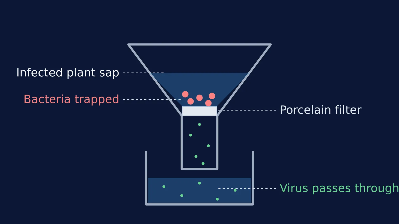
In 1898, the Dutch microbiologist Martinus Beijerinck published a paper showing that the tobacco mosaic agent was fundamentally different from bacteria (Bos, 1999). He demonstrated that the pathogen passed freely through filters designed to trap all known microbes, and that it multiplied only within the dividing tissues of the plant. To describe this strange new entity, he coined the Latin phrase *contagium vivum fluidum*—an infectious living fluid.
What is the contagium vivum fluidum?
The term *contagium vivum fluidum* translates to "infectious living fluid," a concept Beijerinck introduced to explain a pathogen that behaved entirely like a liquid rather than a solid cell.
His conclusion came from a pivotal filtration experiment. Six years earlier, in 1892, the Russian scientist Dmitri Ivanovsky had observed that the sap from infected tobacco leaves remained infectious even after passing through a Chamberland porcelain filter, which possessed pores small enough to catch any known bacteria (Bos, 1999). Ivanovsky, constrained by the existing germ theory, assumed the filter was flawed or that the disease was simply caused by a secreted bacterial toxin. He kept looking for a conventional microbe.
Beijerinck repeated the filtration and went a crucial step further to test the toxin hypothesis. He took the filtered, bacteria-free sap and used it to infect a healthy tobacco plant. Once that plant developed the mosaic disease, he extracted its sap, filtered it, and infected a third plant. He repeated this chain multiple times, showing that the infectious agent did not run out or become diluted. It was actively multiplying, proving it was a living pathogen and not merely a finite chemical toxin. Because the agent passed through the fine filter, multiplied in close association with the host's metabolism and was distributed in the plant's phloem vessels, Beijerinck concluded it was a living fluid.
How did Beijerinck change biological classification?
Before Beijerinck's experiments, biologists operated under the assumption that all infectious diseases were caused by cellular microorganisms that could be cultured independently on artificial nutrient media, such as agar plates or meat broths.
Beijerinck found that the tobacco mosaic agent openly defied this rule. No matter what combination of nutrients he provided, the pathogen refused to grow in a laboratory dish. It only multiplied when in direct contact with the dividing, living cells of a host plant. This observation established the concept of obligate parasitism for these new agents: they require a host's living cellular machinery to reproduce.
While Beijerinck's belief that the pathogen was a true, structureless fluid was eventually corrected—W. M. Stanley proved in 1935 that the tobacco mosaic virus could be isolated and crystallized as discrete, protein-based particles (Bos, 1999)—his 1898 paper laid the conceptual foundation for virology. He was the first to recognize that viruses represent a completely distinct category of biological entity, operating at the very threshold of life.
How is the history of viruses tested in exams?
The timeline of virology is a common topic in the biological classification chapter of the Class 11 biology syllabus. To prepare for multiple-choice questions on this topic, ensure you can match these three scientists to their exact contributions:
D. J. Ivanovsky (1892): Discovered that the mosaic disease of tobacco was caused by microbes smaller than bacteria, as they passed through bacteria-proof filters.
M. W. Beijerinck (1898): Demonstrated that the extract of infected plants caused infection in healthy plants. He named the pathogen *contagium vivum fluidum*.
W. M. Stanley (1935): Showed that viruses could be crystallized and that these crystals consist largely of proteins.
Understanding how early scientists deduced the nature of viruses without ever seeing them through a microscope is an excellent exercise in scientific reasoning. If you are studying these milestones, the Fermi Concept Library contains dedicated lessons on the discovery and structure of viruses.
When you start a session, Fermi's AI tutor can work out what you already know about the five-kingdom system and build a plan for your study time. Rather than just giving you a list of facts to memorize, it holds a line of reasoning, checking your understanding of concepts like obligate parasitism and non-cellular life as it goes.
For more on how organisms are grouped, read about the viruses, viroids and lichens that sit completely outside the main biological kingdoms, or review the six enzyme classes that drive metabolic reactions in living cells.


