On this page
In the mid-1950s, neuroscientists viewed the brain as an electrical network. Synaptic transmission was widely assumed to operate through direct electrical currents, and chemical signalling was treated with scepticism outside peripheral nerves. When Hermann Blaschko mapped the catecholamine pathway in 1939, he identified dopamine (3-hydroxytyramine) merely as an intermediate between tyrosine and noradrenaline. For nearly two decades, textbooks dismissed dopamine as an inactive metabolic stepping stone with no function of its own.
Why was dopamine long dismissed as an inactive chemical precursor?
This view began to shift through experiments with reserpine, an alkaloid from *Rauwolfia serpentina*—a plant whose active constituents were isolated by chemist Salimuzzaman Siddiqui. In 1955 at the National Institutes of Health, Bernard Brodie showed that reserpine depleted brain serotonin, attributing animal sedation entirely to serotonin loss.
Swedish pharmacologist Arvid Carlsson, who had spent five months in Brodie's laboratory, suspected catecholamines were involved, as he recalled in his Nobel lecture. Collaborating with Nils-Åke Hillarp at the University of Lund, Carlsson found reserpine also depleted catecholamines. When rabbits received reserpine (5 mg/kg intravenously), they developed severe akinesia: loss of spontaneous motion, drooping ears, and hunched rigidity, closely resembling human Parkinson's disease.
How did the 1957 rabbit experiment prove dopamine controls motor function?
To test which amine was responsible, Carlsson, Margit Lindqvist, and Tor Magnusson injected precursors into reserpinised rabbits, reporting their findings in *Nature* in 1957. Injecting 5-hydroxytryptophan (5-HTP) restored brain serotonin, but the animals remained completely paralysed.
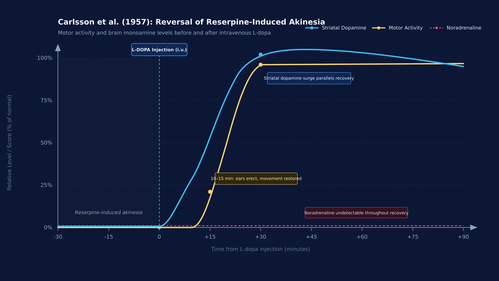
Next, they injected intravenous L-dopa (3,4-dihydroxyphenylalanine), the precursor to dopamine and noradrenaline. Within 10 to 15 minutes, the rabbits woke up, raised their ears, and regained full motor coordination.
Crucially, using a fluorimetric assay described in *Science* in 1958, Carlsson's team showed that brain noradrenaline remained undetectable during this recovery, while dopamine levels surged. In 1959, Åke Bertler and Evald Rosengren published in *Experientia* that approximately 80% of brain dopamine is concentrated in the corpus striatum, a basal ganglia motor centre. Dopamine was thus proven to be an independent motor neurotransmitter, earning Carlsson the 2000 Nobel Prize in Physiology or Medicine, documented in *Bioscience Reports*.
Why does L-dopa cross the blood-brain barrier while dopamine cannot?
If dopamine deficiency causes akinesia, why not administer dopamine directly? Cerebral capillaries are sealed by endothelial tight junctions. At physiological pH, dopamine is positively charged and hydrophilic, preventing it from crossing the lipid membrane.
L-dopa, a neutral amino acid, crosses the blood-brain barrier using the Large Neutral Amino Acid Transporter 1 (LAT1). Inside the brain, aromatic L-amino acid decarboxylase (AADC) converts L-dopa directly into active dopamine.
How did Carlsson's discovery lead to Parkinson's disease therapies?
In 1960, Herbert Ehringer and Oleh Hornykiewicz reported in *Klinische Wochenschrift* that post-mortem striatal samples from Parkinson's patients showed an 80% to 90% deficit in dopamine.
In 1961, Hornykiewicz and Walther Birkmayer administered intravenous L-dopa to bedridden patients, temporarily restoring movement. By 1967, George Cotzias and colleagues demonstrated in the *New England Journal of Medicine* that gradual oral dosing provided sustained control of Parkinsonian symptoms. Today, L-dopa remains the cornerstone of therapy, co-prescribed with peripheral decarboxylase inhibitors to prevent breakdown before entering the brain.
How to analyse neurochemical pathways: a 4-step framework for biology students
Carlsson's logic provides a 4-step framework for evaluating neural signalling and pharmacological interventions:
Map biosynthetic bottlenecks: Identify the substrate, rate-limiting enzyme (such as tyrosine hydroxylase), and immediate product.
Assess membrane permeability: Check molecular charge and transporter mechanisms (such as LAT1 carrier transport versus tight-junction exclusion).
Correlate localisation with anatomy: Connect where the transmitter concentrates to the functional anatomy of the brain, matching striatal pathways with motor control.
Distinguish chemical loss from cellular loss: Differentiate temporary transmitter depletion from degenerative death of neurons, as seen in Alzheimer's disease neurodegeneration.
Mastering these multi-step pathways requires connecting molecular reactions to observable physiology. When mapping biochemical regulation, the Fermi Concept Library covers every concept in physics, chemistry, biology, mathematics and computing, each with its own lesson and its place in the map. When solving physiology problems, the Fermi Problem Library explains step-by-step reasoning rather than printing an answer.