On this page
Albert Claude wanted to know what was inside a cell, and the microscope of his day could not show him. So he took a more direct route: he broke cells open, spun the pieces in a centrifuge, and let gravity, sped up, sort them by size and weight. That method, cell fractionation by differential centrifugation, is why your Class 11 chapter on the cell can say what mitochondria and the endoplasmic reticulum actually do.
Who was Albert Claude?
Claude earned his medical degree from the Université de Liège in Belgium in 1928 and joined the Rockefeller Institute in New York in the summer of 1929, according to his Nobel Prize biography. In his Nobel lecture he remembered student hours at the light microscope, "gazing at the blurred boundary" inside the cell, until he decided he "should stop that futile game, and should try something else."
His first job at Rockefeller was chemical: isolating the agent behind a chicken tumour. Along the way, small particles from the cell's ground substance had "settled in one of my test tubes." In the lecture he calls them microsomes.
In 1974 Claude shared the Nobel Prize in Physiology or Medicine with Christian de Duve and George E. Palade "for their discoveries concerning the structural and functional organization of the cell" (Nobel Prize facts page).
How does cell fractionation by centrifugation work?
The 1974 Nobel press release explains the principle simply. Cells are carefully homogenised and their parts settle towards the bottom of a test tube; a centrifuge speeds up that settling, and the parts come down in steps, heaviest first, starting with the nuclei, then the others in order of weight.
Claude's own description was blunter. His fragments were "obtained by rubbing cells in a mortar" and put through "multiple cycles of sedimentations, washings and resuspensions" (Nobel lecture). The pieces kept working in the test tube as they would inside the cell. He set out the method in two papers in 1946, the first being Fractionation of mammalian liver cells by differential centrifugation in the Journal of Experimental Medicine.
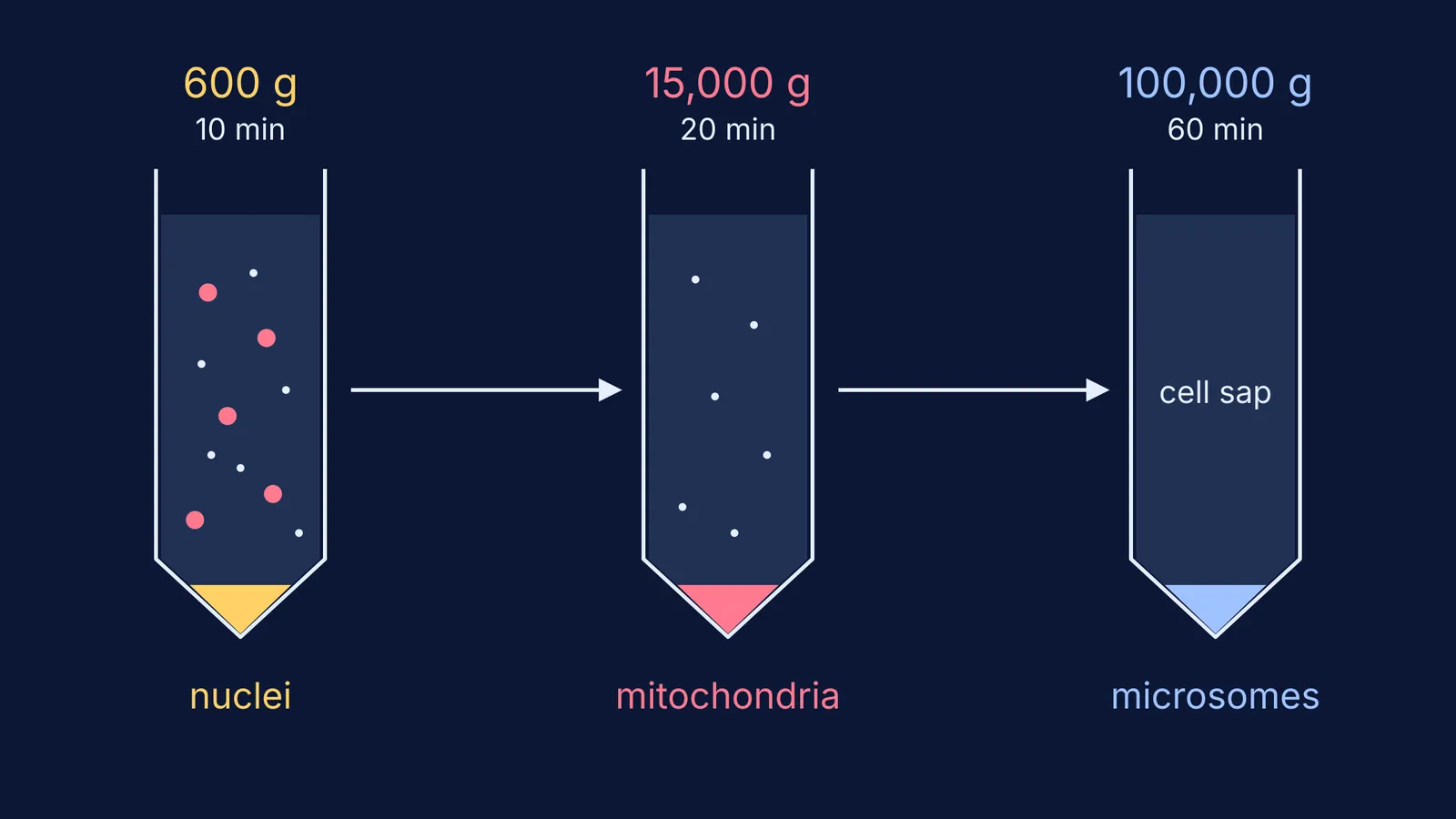
Here is the sequence with typical modern settings, from the table of typical parameters in Wikipedia's article on differential centrifugation. Exact values vary by tissue and protocol. The g number says how many times Earth's gravity the spin applies.
Spin the broken-cell mixture at about 600 g for 10 minutes. Nuclei collect as a pellet at the bottom.
Pour off the liquid above (the supernatant) and spin it at about 15,000 g for 20 minutes. Mitochondria pellet, with lysosomes, peroxisomes and, in plant cells, chloroplasts.
Spin that supernatant at 50,000 to 100,000 g for 60 minutes. The microsomal fraction, pieces of endoplasmic reticulum and plasma membrane, pellets.
What is left is the soluble part of the cytoplasm, which Claude's generation called cell sap.
What did Claude find inside the fractions?
The four fractions from Claude's procedure were nuclei, mitochondria, microsomes and cell sap, says the Nobel press release. Testing each fraction chemically, Claude and his colleagues showed that key respiratory enzymes, including cytochrome oxidase and cytochrome c, sat in the mitochondria (Nobel lecture). That led him to call mitochondria "the real power plants of the Cell", an early form of the "powerhouse" idea in your textbook. With the electron microscope, his microsomes turned out to be pieces of the endoplasmic reticulum.
De Duve later spun cells the same way, found enzymes that fitted none of the four fractions, and tracked them to a fifth: lysosomes. Read how Christian de Duve found lysosomes and peroxisomes. Palade combined centrifugation with electron microscopy to study ribosomes and secretion, told in George Palade and the ribosomes in the cell's protein factory.
How do you remember the order of cell fractions?
Use one rule: bigger, heavier particles come down at lower g and in less time. Claude's Nobel lecture describes mitochondria as small bodies about half a micron in diameter. A nucleus is much larger, and microsomes are smaller fragments still. So the ladder is nuclei, then mitochondria, then microsomes, then cell sap.
Test yourself:
You skip the 600 g step and spin the whole mixture at 15,000 g. What is in the pellet? Nuclei and mitochondria together, because nothing removed the nuclei first.
You want to test where respiration enzymes sit. Which fraction do you take? The mitochondrial pellet, as Claude did.
A friend says the cell sap is a pellet. What do you say? It is the final supernatant, the liquid left after every spin.
If the order keeps slipping, the Fermi Concept Library gives every biology concept its own lesson and its place in the map, and Fermi's AI tutor checks your understanding as it goes and notices where you are stuck.
For the full organelle list, continue with Cell: the unit of life for NEET, with organelles compared.