

On this page
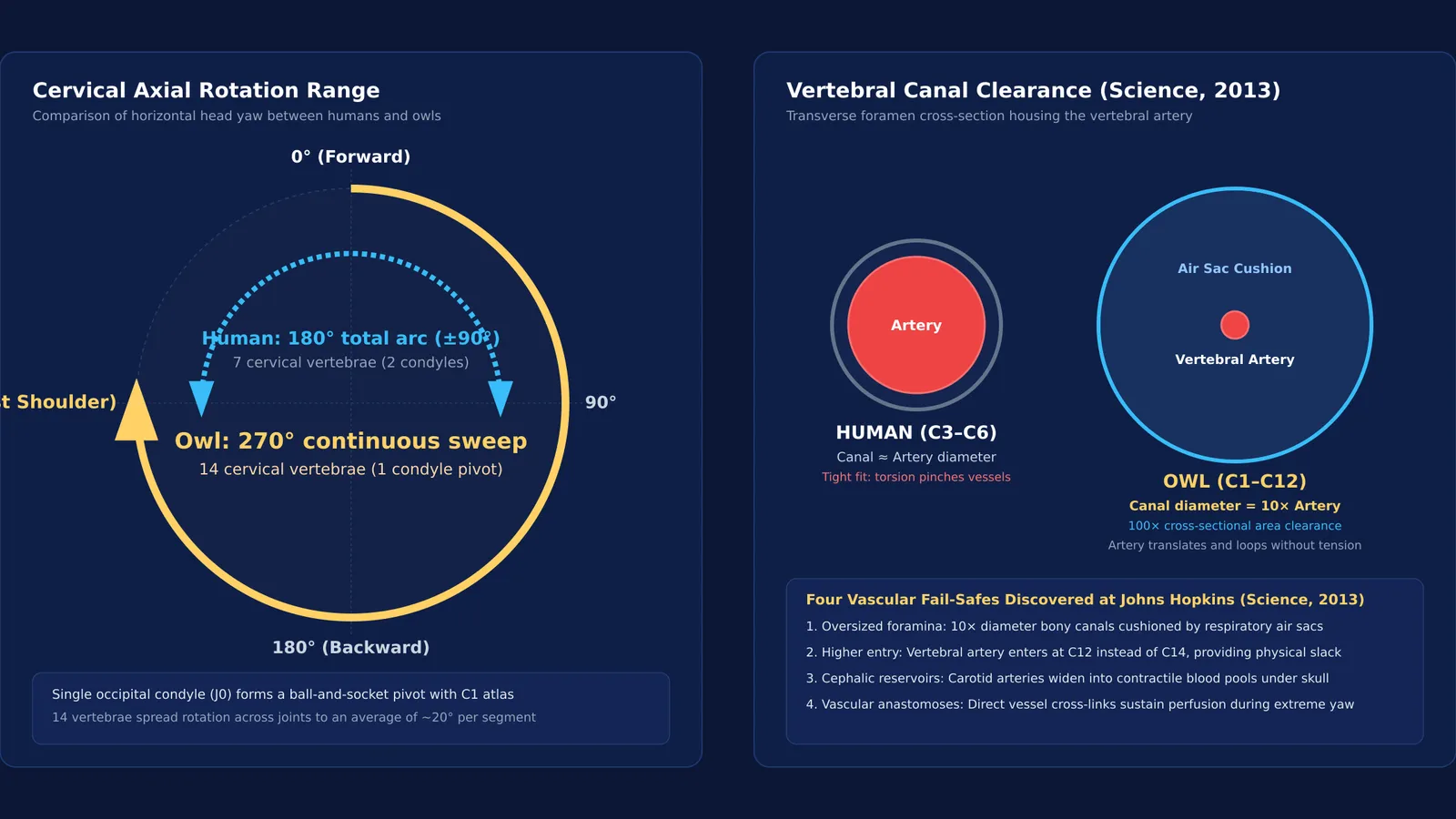
If you try to turn your head over your shoulder, your neck joints stop at roughly 90 degrees in either direction. For humans, forcing rotation beyond that threshold tears arterial linings, pinches nerves, and cuts off cerebral blood supply. Yet an owl perched on a branch can twist its head 270 degrees in one smooth sweep—facing backward and continuing past the opposite shoulder—without shifting its body or suffering vascular damage (Science 2013).
This biomechanical ability is an evolutionary necessity born from an anatomical trade-off between nocturnal vision and skeletal architecture.
Why can't owls move their eyes in their sockets?
Human eyes are spherical globes suspended in soft tissue, steered by six extraocular muscles that let you track moving objects without turning your neck. In contrast, an owl's eyes are elongated cylindrical tubes shaped like binoculars.
This tubular design creates room for a massive cornea and lens relative to skull size, maximizing photon capture in near-total darkness. But that optical advantage carries a mechanical consequence. Each eye is encased inside the skull by a rigid cone of 14 to 15 overlapping bony plates called a sclerotic ring (Veterinary Medicine and Science 2022).
Because these bony rings anchor the eyes firmly to the cranium, owls have virtually zero independent eyeball movement, shifting by less than 2 to 3 degrees. In Vision Research, Graham Martin recorded that an owl's forward-facing eyes yield a total visual field of roughly 110 degrees, with 48 to 70 degrees of binocular overlap for hunting depth perception. That leaves a blind zone of 160 degrees or more directly behind them. Unlike humans, whose optics can be studied through refractive defects of the human eye, an owl cannot glance sideways. To inspect rustling prey, it must swivel its entire head.
How do owls rotate their necks without cutting off blood flow?
In human medicine, sudden neck twists frequently stretch the vertebral and carotid arteries against bone, causing arterial tears, clots, and stroke. How owls twist through 270 degrees without injury remained unclear until 2013, when Dr. Philippe Gailloud and Fabian de Kok-Mercado at Johns Hopkins University mapped owl cranial vasculature using angiography and CT imaging (Science 2013). Their study identified four vascular adaptations:
Oversized transverse foramina: In 12 of the owl's 14 cervical vertebrae, the bony canals housing the vertebral arteries are roughly 10 times wider than the vessels themselves. Cushioning air sacs from the respiratory system fill this space, allowing the arteries to slide without compression.
Higher vascular entry: The vertebral artery enters the spine at the 12th cervical vertebra rather than the 14th, creating mechanical slack along the neck base.
Contractile cephalic reservoirs: Near the skull base, carotid arteries widen into muscular reservoirs that pool oxygenated blood, maintaining brain perfusion even if neck rotation pinches flow below.
Vascular anastomoses: Direct vessel connections link the carotid and vertebral systems, providing collateral bypass pathways that reflect principles seen in body fluids and circulation.
What skeletal adaptations make 270-degree rotation possible?
Arterial protection alone does not produce motion; the cervical skeleton must also provide the physical range. While mammals possess 7 cervical vertebrae, owls have 14 cervical vertebrae, doubling the articulation count found in the human axial skeleton (Journal of Anatomy 2017).
Furthermore, human skulls rest on the spine via two occipital condyles, which stabilize the head but restrict axial yaw. Owls possess only a single occipital condyle, forming a ball-and-socket joint at the atlanto-occipital junction.
Biomechanical research led by Markus Krings and colleagues demonstrated that this cervical column divides into distinct kinematic zones:
The upper cervical joints deliver the primary horizontal yaw.
The middle vertebrae provide postural stability.
The lower cervical joints combine rolling and lateral flexion, shifting the head's center of mass so the bird retains balance during extreme turns.
A student framework: analyzing evolutionary adaptations
By synthesizing morphological data from Science 2013 and the Journal of Anatomy 2017, you can analyze multi-system animal adaptations using a four-step framework:
Sensory driver: Identify the sensory trade-off (tubular eyes maximize nocturnal light capture but require sclerotic rings that eliminate eye movement).
Segmental division: Count the articulating vertebrae (14 cervical vertebrae and their saddle-shaped heterocoelous joints reduce the required rotation to an average of roughly 19 to 21 degrees per joint, preventing excessive shear on any single articulation).
Articulation geometry: Note the joint structure (monocondylic ball-and-socket pivot vs dicondylic hinge).
Hemodynamic fail-safes: Account for continuous fluid flow (10-to-1 canal clearance, contractile reservoirs, and collateral shunts).
Visualizing complex biological mechanics
Understanding how 3D kinematics, fluid dynamics, and bone geometry interact can be challenging from two-dimensional textbook diagrams. The Fermi Simulation Lab provides interactive simulations for ideas that are easier to see than to read, helping you explore spatial mechanics dynamically.
When you need to review vertebrate organ systems across your syllabus, the Fermi Concept Library offers lessons covering every concept in physics, chemistry, biology, mathematics and computing, each placed within its concept map.


