
On this page
Body Fluids and Circulation mixes three kinds of question: numbers (cell counts, cardiac output), sequences (the cardiac cycle) and rules (blood group compatibility). Each has its own trap. This post works through all three with NCERT's values, explains the ECG, and lists the misconceptions that physiology teachers find most often.
What does the syllabus include?
The NEET (UG)-2026 syllabus lists composition of blood, blood groups, coagulation, lymph, the structure of the heart and blood vessels, the cardiac cycle, cardiac output, ECG, double circulation, regulation of cardiac activity, and four disorders. All of it is in NCERT Class 11 Chapter 15. Check the syllabus for your exam year.
What is blood made of?
Plasma: nearly 55 per cent of blood; 90 to 92 per cent water and 6 to 8 per cent proteins. Fibrinogen is for clotting, globulins for defence and albumins for osmotic balance. Plasma without clotting factors is serum.
Formed elements: nearly 45 per cent of blood.
RBCs: 5 to 5.5 million per mm³ in a healthy adult man; 12 to 16 g of haemoglobin per 100 mL; average life 120 days; destroyed in the spleen.
WBCs: 6000 to 8000 per mm³. Neutrophils 60 to 65 per cent, lymphocytes 20 to 25, monocytes 6 to 8, eosinophils 2 to 3, basophils 0.5 to 1.
Platelets: 150,000 to 350,000 per mm³, made from megakaryocytes.
Clotting runs as a cascade: injury triggers thrombokinase, which helps convert prothrombin to thrombin; thrombin converts fibrinogen to fibrin threads. Calcium ions are essential.
How do ABO and Rh groups work?
Karl Landsteiner received the 1930 Nobel Prize "for his discovery of human blood groups". NCERT's table:
Group A: antigen A, anti-B in plasma; can receive from A and O.
Group B: antigen B, anti-A; can receive from B and O.
Group AB: antigens A and B, no antibodies; can receive from all four groups (universal recipient).
Group O: no antigens, both anti-A and anti-B; can receive only from O, but can donate to all (universal donor).
Worked check: a group B patient has anti-A in plasma. Group A or AB blood carries antigen A, so it would clump. Group B and group O blood are safe.
About 80 per cent of people are Rh positive. An Rh-negative mother carrying an Rh-positive foetus can be exposed to foetal blood at the first delivery and start making anti-Rh antibodies, which can harm a later Rh-positive foetus (erythroblastosis foetalis). Giving the mother anti-Rh antibodies right after the first delivery prevents this.
Worked example: cardiac cycle numbers
Cycle length: 72 beats per minute means 60 ÷ 72 = 0.83 seconds per cycle, which NCERT rounds to 0.8 s.
Cardiac output: 70 mL × 72 = 5040 mL per minute, about 5 litres.
During exercise (illustrative): if heart rate rose to 120 and stroke volume to 100 mL, output would be 120 × 100 = 12,000 mL, or 12 litres per minute. NCERT notes that the body can change both stroke volume and heart rate.
From an ECG: if you count 12 QRS complexes in 10 seconds, the heart rate is 12 × 6 = 72 beats per minute.
What happens in each step of the cardiac cycle?
Joint diastole: all four chambers relax; AV valves are open and blood flows into the ventricles. Semilunar valves are closed.
Atrial systole: the SAN fires and both atria contract, adding about 30 per cent more blood to the ventricles.
Ventricular systole: the impulse passes through the AVN, AV bundle and bundle of His. Ventricles contract, AV valves close (the first sound, lub) and semilunar valves open.
Ventricular diastole: pressure falls and semilunar valves close (the second sound, dub). AV valves then open and filling starts again.
The SAN generates 70 to 75 action potentials a minute and is the pacemaker; the heart is myogenic. Sympathetic nerves raise heart rate and output, and parasympathetic nerves lower them.
How do you read a standard ECG?
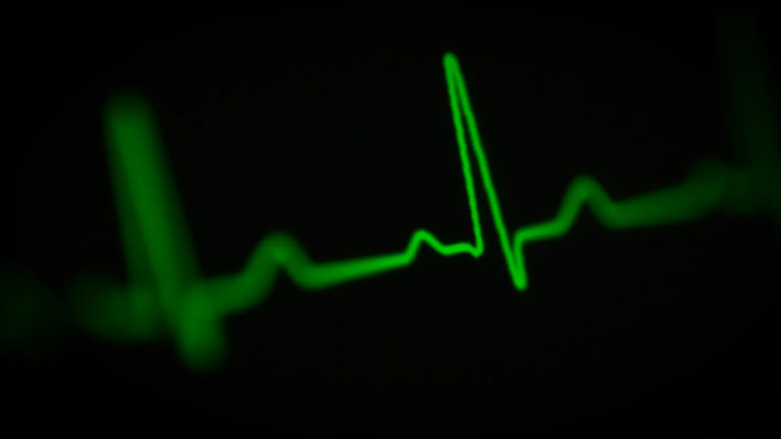
A standard ECG uses three leads, one on each wrist and one on the left ankle. Willem Einthoven received the 1924 Nobel Prize "for his discovery of the mechanism of the electrocardiogram".
P wave: depolarisation of the atria, leading to atrial contraction.
QRS complex: depolarisation of the ventricles. Contraction starts shortly after Q and marks the start of systole.
T wave: repolarisation of the ventricles. The end of T marks the end of systole.
Which misconceptions are common?
Michael and colleagues (2002) surveyed undergraduates in 12 courses at 8 institutions and found 13 cardiovascular misconceptions, each held by 20 to 81 per cent of students. They suggest one source is difficulty applying simple general models to specific cases. Common slips at school level:
Calling all arteries oxygenated. The pulmonary artery carries deoxygenated blood to the lungs.
Mixing up circulation types: fishes have single circulation, amphibians and most reptiles incomplete double circulation, and birds and mammals complete double circulation.
Treating heart failure, cardiac arrest and heart attack as the same. NCERT separates them.
Misreading blood pressure: 120/80 is normal; repeated readings of 140/90 or higher indicate hypertension.
If you understand the cycle but still freeze on numerical questions, the Fermi Problem Library has worked problems with a step-by-step solver that explains the reasoning.
A practice routine
Write the four cardiac-cycle steps from memory, marking valve positions in each.
Draw a standard ECG and label P, QRS and T with what each means.
Do three cardiac output sums with different heart rates and stroke volumes.
Test blood group compatibility for all 16 donor and recipient pairs.
Connect this chapter to breathing and exchange of gases and to the kidney and nephron, which filters about a fifth of the cardiac output.
Today, draw the ECG from memory and write one line under each wave.