
On this page
Excretory Products and their Elimination is short, but almost every line is examinable: which animals excrete what, which segment of the nephron does what, and which hormone raises or lowers blood pressure. The numbers also fit together neatly once you work them through. This post does that, explains the counter-current mechanism in plain steps, and ends with a practice routine.
What does the syllabus include?
The NEET (UG)-2026 syllabus lists modes of excretion, the human excretory system, urine formation, osmoregulation, regulation of kidney function (renin-angiotensin, atrial natriuretic factor, ADH and diabetes insipidus), the role of other organs, disorders, and dialysis. The chapter is NCERT Class 11 Chapter 16. Check the syllabus for your exam year.
Which animals excrete ammonia, urea or uric acid?
Ammonotelic: ammonia is the most toxic and needs the most water. Many bony fishes, aquatic amphibians and aquatic insects; ammonia leaves by diffusion across body or gill surfaces.
Ureotelic: mammals, many terrestrial amphibians and marine fishes. The liver converts ammonia to urea.
Uricotelic: uric acid is the least toxic and needs the least water. Reptiles, birds, land snails and insects.
Excretory structures: protonephridia or flame cells (flatworms, rotifers, some annelids, *Amphioxus*), nephridia (earthworms), Malpighian tubules (most insects) and antennal or green glands (prawns).
What are the parts of a nephron?
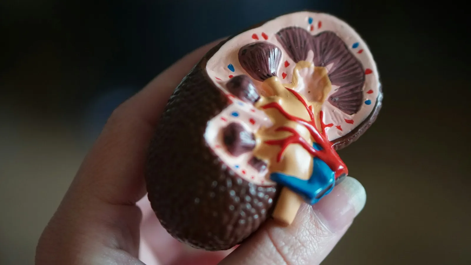
Each kidney has about a million nephrons. A nephron is a glomerulus, a tuft of capillaries fed by the afferent arteriole and drained by the efferent arteriole, inside a Bowman's capsule. Together they form the Malpighian body. The tubule continues as the PCT, the hairpin loop of Henle, the DCT and a collecting duct. The Malpighian body, PCT and DCT lie in the cortex; the loop dips into the medulla. Most nephrons are cortical, with short loops. Juxtamedullary nephrons have long loops and a U-shaped capillary alongside, the vasa recta.
Worked example: the numbers in urine formation
Daily filtrate: GFR is about 125 mL per minute. 125 × 60 × 24 = 180,000 mL, or 180 litres a day.
Reabsorption: urine is about 1.5 litres a day, so 180 − 1.5 = 178.5 litres are reabsorbed. 178.5 ÷ 180 = 0.992, or about 99 per cent.
Share of the heart's output: the kidneys filter 1100 to 1200 mL of blood a minute. Against a cardiac output of about 5000 mL a minute, 1200 ÷ 5000 = 0.24, which NCERT describes as roughly one-fifth.
Concentration: the gradient runs from 300 mOsmol per litre in the cortex to about 1200 in the inner medulla. 1200 ÷ 300 = 4, so urine can be about four times as concentrated as the initial filtrate.
What does each part of the tubule do?
PCT: brush-border epithelium. Reabsorbs nearly all essential nutrients and 70 to 80 per cent of electrolytes and water. Secretes H+ and ammonia; absorbs HCO3−.
Descending limb of Henle: permeable to water, almost impermeable to electrolytes. The filtrate becomes more concentrated as it goes down.
Ascending limb: impermeable to water; electrolytes move out. The filtrate becomes more dilute as it goes up.
DCT: conditional reabsorption of Na+ and water; secretes H+, K+ and NH3.
Collecting duct: can reabsorb large amounts of water. Lets small amounts of urea into the medulla, and secretes H+ and K+.
How does the counter-current mechanism work?
Filtrate flows down one limb of the loop and up the other, in opposite directions. Blood in the two limbs of the vasa recta also flows in opposite directions.
The ascending limb moves NaCl into the medulla. The descending limb of the vasa recta picks it up, and the ascending vasa recta returns it to the interstitium.
Small amounts of urea enter the thin ascending limb and are returned to the interstitium by the collecting tubule.
NaCl and urea together build the gradient from 300 to about 1200 mOsmol per litre.
As the collecting duct passes through this salty medulla, water leaves it, concentrating the urine.
How is kidney function regulated?
ADH: loss of body fluid activates osmoreceptors; the hypothalamus releases ADH (vasopressin) from the neurohypophysis. ADH increases water reabsorption from the later parts of the tubule, reducing urine output.
Renin-angiotensin: a fall in GFR makes JG cells release renin. Angiotensinogen becomes angiotensin I and then angiotensin II, a vasoconstrictor that raises glomerular pressure and triggers aldosterone release, increasing Na+ and water reabsorption.
ANF: more blood flow to the atria releases atrial natriuretic factor, which dilates vessels and lowers blood pressure, acting as a check on renin-angiotensin.
Other facts: urine is slightly acidic, about pH 6; adults excrete 25 to 30 g of urea a day; the lungs remove about 200 mL of CO2 a minute. Glucose or ketone bodies in urine indicate diabetes mellitus. In haemodialysis, heparin is added before the blood enters the dialyser and anti-heparin before it returns.
Why test yourself instead of rereading?
Roediger and Karpicke (2006) had students either reread prose passages or take recall tests on them. Rereading looked better after five minutes, but on tests two days and one week later, the students who had practised recall remembered substantially more, even though rereading had made the other group more confident.
For this chapter, that means drawing a blank nephron and filling in each segment's job from memory. If you are not sure where to start across the physiology unit, Fermi's AI tutor can work out what you already know and build a plan for the session.
A practice routine
Draw a nephron with vasa recta and label every part without notes.
Redo the four calculations above from memory.
Write the three hormone pathways as arrow chains.
Turn the true-or-false items in NCERT's exercises into flashcards that test recall.
Revise body fluids and circulation alongside this chapter, since the two share blood pressure and cardiac output.
Today, draw the loop of Henle and mark where water leaves and where salt leaves.