

On this page
If you wake up two mornings after a sports match, cricket practice, or your first gym workout feeling painfully stiff, you are experiencing delayed onset muscle soreness (DOMS). While common locker-room advice blames "lactic acid settling in your muscles," exercise physiology tells a very different story. Next-day soreness has nothing to do with lactic acid. Instead, it stems from microscopic mechanical damage to the contractile units inside your muscle fibres and the delayed inflammatory healing process that follows.
Why do muscles ache a day after exercise instead of right away?
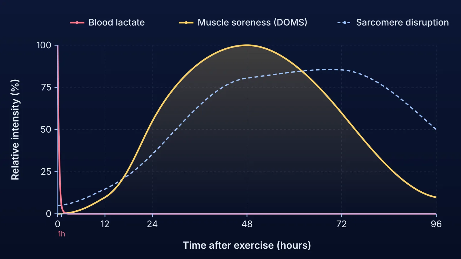
Acute fatigue strikes during heavy exertion and fades within an hour as breathing slows and cellular energy stores replenish. Soreness, however, follows a curiously delayed schedule.
This delay has been documented for over a century. In 1902, researcher Theodore Hough published the first ergographic study of muscular soreness in the American Journal of Physiology. Hough separated immediate muscular fatigue from the soreness that developed 8 to 24 hours later, concluding that next-day pain was "fundamentally the result of ruptures within the muscle" rather than metabolic waste. A comprehensive review by Cheung, Hume, and Maxwell (2003) confirmed this timeline: DOMS typically emerges 12 to 24 hours after unfamiliar exercise, peaks between 24 and 72 hours, and resolves within 5 to 7 days.
Why is lactic acid not the cause of delayed muscle soreness?
The myth persists because intense exertion does produce lactate. When contracting muscle demands ATP faster than aerobic respiration can supply it, anaerobic glycolysis splits glucose into pyruvate and converts it to lactate to regenerate NAD⁺, as traced in Otto Meyerhof's discovery of lactic acid in glycolysis.
However, lactate is rapidly cleared. Research by Menzies et al. (2010) demonstrated that blood lactate returns to resting baseline concentrations within 30 to 60 minutes after exhaustive exercise. The liver converts lactate back into glucose via the Cori cycle, while heart muscle and resting skeletal fibres oxidise it directly for fuel.
The decisive disproof came from Schwane et al. (1983). Runners performed two treadmill bouts: flat running and running down a 10% incline. Flat running demanded high metabolic effort, spiking blood lactate, yet caused no delayed soreness over the next 72 hours. Downhill running demanded far less metabolic energy and produced no lactate spike, yet triggered severe DOMS.
For students who need to master the steps of cellular respiration and anaerobic fermentation, the Fermi Concept Library holds every concept in physics, chemistry, biology, mathematics and computing, each with its own lesson and its place in the map.
How do eccentric contractions disrupt sarcomeres and Z-discs?
Downhill running causes severe soreness because it relies on eccentric contractions—movements where active muscle fibres lengthen under load, acting as mechanical brakes. Because fewer motor units are recruited to resist a load than to lift it, the mechanical stress per active muscle fibre is far higher.
In skeletal muscle, the contractile unit is the sarcomere, bounded by Z-discs, as detailed in our guide to locomotion and movement and how calcium and cross-bridges drive muscle contraction. During eccentric lengthening, sarcomeres stretch unevenly. As Proske and Morgan (2001) showed in their "popping sarcomere" model, the weakest sarcomeres stretch beyond actin-myosin filament overlap and "pop," transferring mechanical overload onto neighbouring structures and tearing structural proteins.
Direct microscopic proof was documented by Fridén, Sjöström, and Ekblom (1983). Biopsies taken after eccentric exercise revealed marked Z-band streaming, broadening, and disruption in 30% to 50% of muscle fibres 3 days post-exercise, with fast-twitch Type 2 fibres suffering the greatest damage.
This structural disruption triggers a delayed biochemical chain reaction. Damaged membranes allow calcium to leak into the cytoplasm, activating proteases that degrade cellular components. Over the following days, invading immune cells release prostaglandins and histamines that cause localized swelling and sensitise polymodal nociceptors (pain receptors). It is this delayed inflammation, rather than acute metabolic stress, that causes the tender ache.
If you want to visualise how thin actin and thick myosin filaments interact under mechanical stress, the Fermi Simulation Lab provides interactive simulations for the ideas that are easier to see than to read.
What actually relieves DOMS, and how do muscles adapt?
Because DOMS is driven by tissue repair, passive waiting is rarely the most comfortable option. A practical recovery routine includes:
Active recovery: Light movement like walking or cycling temporarily increases local blood flow and reduces stiffness without further tearing muscle fibres.
Nutritional support: Adequate protein intake supplies amino acids to rebuild torn Z-discs and contractile proteins.
Gentle mobility over aggressive stretching: Forcing an already torn muscle into deep static stretches can aggravate micro-damage. Gentle joint rotations and easy movements are far safer.
Gradual progression: Progressively overloading weights and running distances prevents overwhelming structural strain.
The upside of DOMS is that your muscles adapt rapidly. As reviewed by McHugh (2003), a single eccentric session triggers the "repeated bout effect." Muscle fibres repair themselves by adding sarcomeres in series along myofibrils. This longitudinal addition reduces strain on individual sarcomeres during future workouts, making you far more resilient the next time you train.


