On this page
Most questions on Locomotion and Movement come down to one picture: a sarcomere, with its bands and lines, before and after contraction. If you can draw it and say which parts change length, you can answer most of them. This post explains why the bands behave as they do, works through a band-length problem with numbers, and covers the skeleton and joints.
What does the syllabus include?
The NEET (UG) 2026 syllabus lists types of movement (ciliary, flagellar, muscular), skeletal muscle with its contractile proteins and contraction, the skeletal system, joints, and six disorders: myasthenia gravis, tetany, muscular dystrophy, arthritis, osteoporosis and gout. The matching chapter is Chapter 17 of NCERT Class 11 Biology. Check the syllabus for your exam year.
What is a sarcomere made of?
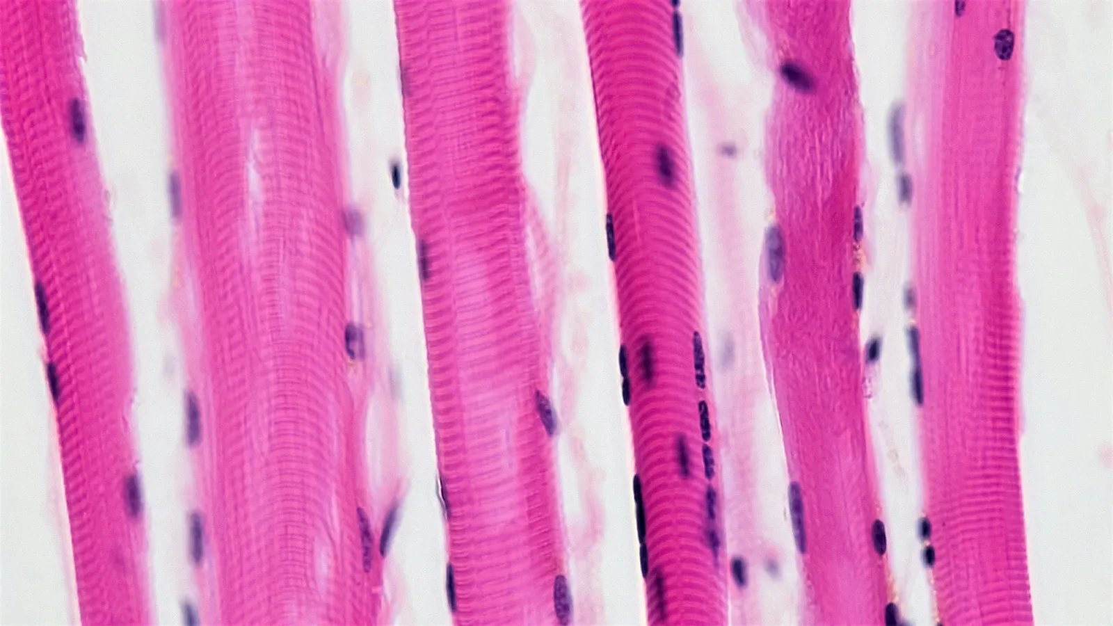
Following NCERT: each muscle fibre holds many myofibrils, and each myofibril is a chain of sarcomeres, the unit of contraction between two Z lines.
Thin filaments are actin, made of two F-actin strands, each a polymer of G-actin, with tropomyosin running alongside and troponin at intervals. They are fixed to the Z line.
Thick filaments are myosin, built from meromyosin units. The head (heavy meromyosin) is an ATPase with binding sites for ATP and actin.
The A band (dark) is the length of the thick filaments. The I band (light) holds only thin filaments and is bisected by the Z line. The H zone is the centre of the A band where thin filaments do not reach. The M line holds the thick filaments together in the middle.
How was the sliding filament theory discovered?
Before the 1950s, as the Biophysical Society's history of the discovery puts it, it was widely accepted that contraction came from myosin filaments shortening. On the same day in 1954, Nature published two papers that pointed to sliding instead: Andrew Huxley and Rolf Niedergerke, using interference microscopy on living frog fibres, and Hugh Huxley and Jean Hanson, using isolated myofibrils. As a historical review by John Squire (2016) explains, the key observation was that as sarcomere length changed, the length of the A band stayed almost constant.
If the A band does not change, the thick filaments do not shorten. The thin filaments must slide past them.
What happens step by step during contraction?
A motor neuron releases acetylcholine at the neuromuscular junction, which sets off an action potential in the sarcolemma.
Calcium ions are released from the sarcoplasmic reticulum into the sarcoplasm.
Calcium binds to troponin, which unmasks the myosin-binding sites on actin.
Using energy from ATP hydrolysis, the myosin head binds actin, forming a cross bridge, and pulls the thin filament towards the centre of the A band.
A new ATP binds, the cross bridge breaks, and the cycle repeats.
When calcium is pumped back, the sites are masked again and the muscle relaxes.
Worked example: which bands shrink?
Use round figures from Squire's review: thick filaments about 1.6 µm long and each set of thin filaments about 1.0 µm long. Ignore the small width of the Z line.
Question. A sarcomere shortens from 2.4 µm to 2.0 µm. Find the A band, total I band and H zone before and after.
A band = thick filament length = 1.6 µm, both before and after.
I band = sarcomere length − A band. Before: 2.4 − 1.6 = 0.8 µm (0.4 µm on each side). After: 2.0 − 1.6 = 0.4 µm.
H zone = sarcomere length − 2 × thin filament length. Before: 2.4 − 2.0 = 0.4 µm. After: 2.0 − 2.0 = 0, so the thin filaments now meet in the middle.
Check: the sarcomere lost 0.4 µm. The I band lost 0.4 µm and the H zone lost 0.4 µm. The A band lost nothing.
This also explains force. Gordon, Huxley and Julian (1966) found that tension in frog fibres peaks on a plateau between sarcomere lengths of 2.05 and 2.2 µm, and said many features of the length-tension curve are simply explained by the sliding filament theory: overlap between thick and thin filaments sets how many cross bridges can pull.
What should you know about the skeleton and joints?
NCERT's counts are standard exam material: 206 bones, with 80 in the axial skeleton; 22 skull bones (8 cranial, 14 facial); 26 vertebrae (7 cervical, 12 thoracic, 5 lumbar, 1 sacral, 1 coccygeal); 12 pairs of ribs, of which 7 are true, 3 false and 2 floating; and 30 bones in each limb.
Joints come in three structural types. Fibrous joints, such as skull sutures, allow no movement. Cartilaginous joints, such as those between vertebrae, allow limited movement. Synovial joints have a fluid-filled cavity and allow a lot. Learn the five synovial examples as pairs: ball and socket (humerus and pectoral girdle), hinge (knee), pivot (atlas and axis), gliding (between carpals) and saddle (carpal and metacarpal of the thumb).
Which mistakes cost marks?
Saying the A band shortens. It stays constant; the I band and H zone shorten.
Putting calcium on tropomyosin. Calcium binds troponin.
Mixing up red and white fibres. Red fibres have more myoglobin and mitochondria and are aerobic; white fibres have more sarcoplasmic reticulum and depend on anaerobic metabolism.
Confusing gout with arthritis. Gout is joint inflammation from uric acid crystals; arthritis is joint inflammation in general.
The signal that starts contraction is covered in our post on neural control and coordination.
A practice routine
Draw a relaxed and a contracted sarcomere side by side and label A, I, H, Z and M.
Do two band-length problems with your own numbers and check that the losses match.
Write the six contraction steps from memory.
Quiz yourself on the skeletal counts and joint pairs after two days, then after a week.
Sliding is easier to see than to read about. The Fermi Simulation Lab has interactive simulations for ideas that are easier to see than read, and the Fermi Concept Library has a lesson for each concept if one step will not stick.
Today, draw the two sarcomeres and write the length of every band on each.