

On this page
When you examine a starfish (*Asterias*) or sea urchin (*Echinus*), you encounter a unique hydraulic engine. As outlined in NCERT Class 11 Biology, Phylum Echinodermata features an endoskeleton of calcareous ossicles and an ambulacral water vascular system. This modified coelomic compartment arises embryonically from the left hydrocoel. Rather than pumping blood through a closed circuit with a heart, echinoderms circulate seawater to power movement, capture prey, and exchange gases.
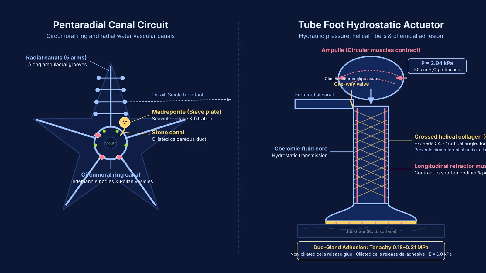
The fluid circuit follows a strict anatomical route:
Madreporite: A porous plate on the aboral surface that admits and filters incoming seawater.
Stone canal: A ciliated duct carrying fluid downward to the central ring.
Ring canal: A circumoral ring circling the mouth, flanked by Polian vesicles for fluid storage and Tiedemann's bodies that form coelomocytes.
Radial canals: Five primary canals extending along the ambulacral grooves of each arm.
Lateral canals: Short side branches with one-way valves that deliver fluid into individual tube feet.
Examinations regularly test this system against the poriferan water canal network, a distinction detailed in our guide to reading NCERT lines with precision. For comparative anatomy across phyla, the Fermi Concept Library covers every concept in biology, each with its own lesson and its place in the map.
How do tube feet generate grip and movement without suction?
Each tube foot is an independent hydrostatic unit consisting of an internal muscular bulb (the ampulla) and an external tubular stem (the podium) ending in a terminal disc. When circular muscles in the ampulla contract, fluid is forced downward into the podium. The lateral canal's one-way valve snaps shut, isolating podial fluid from the radial canal.
In physiological measurements published in the *Journal of the Marine Biological Association of the United Kingdom*, John Binyon (1964) showed that internal hydrostatic pressure peaks at roughly 30 cm H₂O (2.94 kPa) during protraction, dropping to zero at rest. To prevent the soft podium from ballooning into a sphere under pressure, R. S. McCurley and W. M. Kier (1995) revealed in *The Biological Bulletin* that the podial wall is reinforced by a crossed-fiber helical array of collagen fibers oriented at roughly 67°. Because this angle exceeds the critical angle of 54.7°, internal pressure converts into longitudinal extension while resisting circumferential dilation.
Attachment does not depend on simple suction cups. In the *Journal of Experimental Biology*, Romana Santos and colleagues (2005) recorded an elastic modulus of 6.0 kPa for the tube foot disc of *Asterias rubens*, enabling it to conform viscoelastically to substrate micro-roughness. Furthermore, Patrick Flammang and colleagues (1998) demonstrated that sea stars utilize a duo-gland adhesive system. Non-ciliated secretory cells release adhesive proteins that achieve tenacities between 0.18 and 0.21 MPa. Ciliated cells then release de-adhesive secretions to detach, leaving a microscopic footprint.
To pull forward, longitudinal muscles in the podium contract, drawing the animal toward the anchored disc while pushing fluid back into the ampulla.
How do tube feet drive feeding and respiration?
Locomotion is only one function of this hydraulic network. In predatory sea stars, tube feet act as tireless mechanical clamps. When attacking bivalves, the sea star arches over the shell and anchors hundreds of tube feet to both valves. While the mollusc's adductor muscles eventually fatigue under continuous strain, the starfish sustains isometric tension hydraulically with minimal energy expenditure. In *The Biological Bulletin*, Marcel E. Lavoie (1956) demonstrated that a shell gap of just 0.1 mm lets the sea star evert its cardiac stomach into the shell to digest tissues externally.
In suspension feeders such as brittle stars and crinoids, ciliated tube feet trap plankton in mucus and transfer food particles along ambulacral grooves to the mouth.
Simultaneously, tube feet serve as primary respiratory structures. The thin epithelial walls of hundreds of podia provide extensive surface area for oxygen and carbon dioxide diffusion between seawater and coelomic fluid. As highlighted in our guide to animal kingdom classification, echinoderms lack an excretory system. Nitrogenous waste, chiefly ammonia, diffuses directly out of the tube feet into the surrounding water.
Why do radially symmetrical adults develop from bilateral larvae?
Echinoderms are deuterostomes sharing common ancestry with chordates. As described in NCERT Class 11 Biology, their free-swimming larvae (such as bipinnaria and brachiolaria) are bilaterally symmetrical. During metamorphosis, the left coelomic sacs expand to form the water vascular ring and radial canals, while the right side regresses.
This transition yields secondary pentamerous radial symmetry. For slow-moving, benthic animals without a specialized head, radial symmetry allows sensory tube feet to monitor food, physical forces, and chemical cues equally from all directions around the body axis.
Mastering these anatomical transitions requires a systematic approach, such as drawing and labeling NCERT biology diagrams from memory. Apply this 4-step checklist to echinoderm questions:
Identify the canal system: Distinguish the water vascular system with tube feet (Echinodermata) from the choanocyte-lined water canal system (Porifera).
Distinguish symmetry stages: Separate the primary bilateral symmetry of free-swimming larvae from the secondary pentaradial symmetry of adults.
Trace fluid circulation: Follow water from madreporite through stone canal, ring canal, radial canals, lateral canals, and ampullae.
Recall absent systems: Remember that echinoderms lack specialised excretory organs; thin podial walls carry out excretion and gas exchange by diffusion.
For students reviewing physiological mechanisms, the Fermi Problem Library provides worked problems with a step-by-step solver that explains the reasoning rather than printing an answer.


