
On this page
Suck hard on a stiff straw and it stays round. Pinch a soft rubber tube and it folds flat. Your windpipe has to act like the straw when you breathe and give way like the rubber tube when you swallow. The C-shaped rings are how one tube does both, and they explain a single line in your Class 11 NCERT chapter on breathing.
What is the trachea made of?
The trachea is the tube that carries air from the larynx down to the lungs. NCERT describes it as "a straight tube extending up to the mid-thoracic cavity, which divides at the level of 5th thoracic vertebra into a right and left primary bronchi", and says the trachea, bronchi and initial bronchioles "are supported by incomplete cartilaginous rings" (NCERT Class 11 Biology, Chapter 14).
The adult trachea is about 10 to 13 cm long, up from about 5 to 6 cm in infants (StatPearls: Anatomy, Head and Neck, Trachea). It begins just below the larynx and ends at the carina, where it splits into the two bronchi.
Its wall has three parts (OpenStax Anatomy and Physiology 2e, section 22.1):
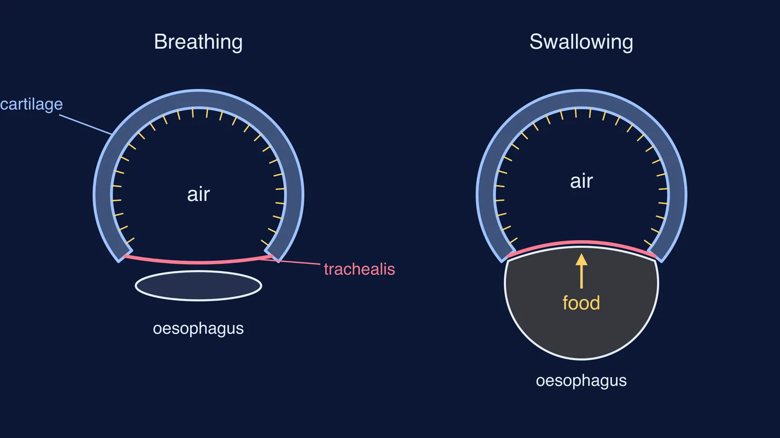
Cartilage rings: 16 to 20 stacked, C-shaped pieces of hyaline cartilage joined by dense connective tissue.
The back wall: the trachealis muscle plus elastic connective tissue, together called the fibroelastic membrane, closing the gap between the two ends of each C.
The lining: pseudostratified ciliated columnar epithelium, continuous with the lining of the larynx.
Our explainer on how cartilage differs from bone places hyaline cartilage in the body.
Why are the tracheal rings C-shaped and not complete circles?
Because the trachea has two jobs, and each part of the ring handles one.
The cartilage stops the airway collapsing. A soft tube can fold flat when air is pulled through it fast. The rings of cartilage "provide structural support and prevent the trachea from collapsing", while the fibroelastic membrane lets the trachea stretch and expand slightly as you breathe in and out (OpenStax).
The open back makes room for food. The oesophagus lies directly behind the trachea, and the structure of its wall makes the trachea "sufficiently flexible and elastic to permit the transient expansion of the esophagus during swallowing" (Kenhub: the trachea). If the rings were full circles, each mouthful would press against hard cartilage; the soft back wall gives way instead.
Keeping food out of the airway altogether is a different structure's job: see how the epiglottis stops food entering the windpipe.
What do the mucus and cilia in the trachea do?
Every breath carries dust and microbes, and the lining traps and clears them. Ciliated cells have "long motile cilia that propel particulate matter entrapped in mucus toward the oropharynx, a process known as mucociliary clearance" (StatPearls).
A review by Ximena Bustamante-Marin and Lawrence Ostrowski gives the numbers (Cilia and Mucociliary Clearance, Cold Spring Harbor Perspectives in Biology, 2017):
About 60% of the cells lining the trachea are ciliated and about 20% are mucus-making goblet cells.
Each mature ciliated cell carries more than 100 cilia, each about 6.5 to 7 µm long.
The cilia beat 10 to 20 times a second.
In the trachea, the mucus layer riding on the cilia is 2 to 5 µm thick.
Beating at that rate, the cilia move mucus out of the airways at about 5.5 mm per minute on average.
For how cilia beat at all, see the 9+2 arrangement inside cilia and flagella.
How long does trapped dust take to leave the trachea?
Take a trachea 11 cm long, inside the 10 to 13 cm adult range from StatPearls, and the clearance speed of 5.5 mm per minute from the mucociliary clearance review.
Convert to one unit: 11 cm = 110 mm.
Divide distance by speed: 110 ÷ 5.5 = 20 minutes.
Check the ends of the range: 100 ÷ 5.5 ≈ 18 minutes and 130 ÷ 5.5 ≈ 24 minutes.
So a speck caught near the bottom reaches your throat in roughly 20 minutes. This uses an average speed; real clearance varies between people.
How do you remember it for the exam?
Use three words: Rings, Room, Rinse.
Rings: C-shaped hyaline cartilage, 16 to 20 of them. Job: stop the collapse.
Room: open back closed by the trachealis muscle and fibroelastic membrane. Job: let the oesophagus expand during swallowing.
Rinse: ciliated lining with goblet cells. Job: mucus traps dust, cilia sweep it up to the throat.
Mistakes to avoid in a written answer:
Calling it elastic cartilage or fibrocartilage. It is hyaline.
Putting the gap at the front. It faces back, towards the oesophagus.
Saying cilia push mucus down to the lungs. They move it up.
Test yourself: draw a cross-section from memory in one minute and label the cartilage, trachealis, oesophagus and cilia. If "pseudostratified" or "hyaline" is where you stall, the Fermi Concept Library has every concept in biology with its own lesson and its place in the map, so you can trace the trachea back to epithelium and cartilage.