

On this page
Think of your digestive system as a long, continuous tube stretching from your mouth to your anus. If food could move freely in both directions, digestion would be chaotic and dangerous—stomach acid would constantly spill into your throat, and waste would travel backwards.
To keep everything moving in one direction, the body uses ring-like bands of muscle called sphincters. These act as one-way valves, opening to let food pass and tightly closing behind it to ensure a smooth, unidirectional flow of digestion.
What are the sphincter muscles in the digestive system?
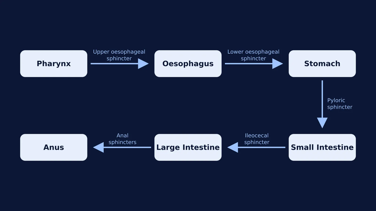
The human digestive tract relies on a series of these valves to separate different chemical environments. According to the standard anatomical definition of sphincters, the main digestive sphincters include:
Upper oesophageal sphincter: Located at the top of the oesophagus, it opens to allow swallowed food to leave the pharynx and enter the oesophagus.
Lower oesophageal sphincter: Also known as the cardiac sphincter, it is found at the junction of the oesophagus and the stomach. It prevents acidic stomach contents from splashing back up.
Pyloric sphincter: Located at the bottom of the stomach, this tough muscle regulates how fast partially digested food (chyme) empties into the duodenum of the small intestine.
Ileocecal sphincter: Situated where the small intestine meets the large intestine, it stops the bacteria-rich contents of the colon from flowing backwards into the ileum.
Anal sphincters: The internal and external anal sphincters are located at the very end of the tract, controlling the exit of waste from the body.
What is the function of the lower oesophageal sphincter?
The lower oesophageal sphincter has a particularly demanding job. The stomach is a highly acidic environment, designed to break down proteins and kill harmful bacteria. The thick lining of the stomach can handle this acid easily, but the delicate tissue of the oesophagus cannot.
When you swallow, this sphincter briefly relaxes to let food drop into the stomach. Immediately afterwards, it must clamp shut. This tight seal is what allows the stomach to churn its contents vigorously without splashing corrosive acid upwards. Without this protective mechanism, the acid meant to digest your food would cause significant damage to the upper digestive tract.
Why does acid reflux happen?
If the lower oesophageal sphincter becomes weak or relaxes at the wrong time, the seal is broken. Stomach contents, including strong digestive acids, escape upwards into the oesophagus. This creates the burning sensation behind the breastbone commonly known as heartburn.
When this occasional reflux becomes a chronic, severe condition, it is called gastroesophageal reflux disease (GERD). The condition is incredibly common. According to the National Institute of Diabetes and Digestive and Kidney Diseases, researchers estimate that about 20% of people in the United States have GERD.
Over time, untreated GERD can cause inflammation in the oesophagus, lead to swallowing difficulties, or even alter the tissue lining the oesophagus.
How can you map the digestive sphincters for exams?
Questions about the digestive system, much like those asking you to map parts of the human brain, appear frequently in biology exams. Because the names of the sphincters tell you exactly where they are, the easiest way to memorise them is to track the journey of food.
If you are stuck on a test question, quickly sketch out this sequence. Just as you might divide the human skeleton into axial and appendicular bones to make it easier to remember, grouping the digestive tract by its organs and valves makes the whole system intuitive:
Pharynx to oesophagus: You need a valve at the top of the oesophagus. That is the upper oesophageal sphincter.
Oesophagus to stomach: You need a valve at the bottom of the oesophagus. That is the lower oesophageal sphincter.
Stomach to small intestine: The lower part of the stomach is called the pylorus, so the valve here is the pyloric sphincter.
Small intestine to large intestine: The last part of the small intestine is the ileum, and the first part of the large intestine is the cecum. Therefore, the valve is the ileocecal sphincter.
Large intestine to the outside: These are the anal sphincters.
If you are preparing for exams and keep confusing anatomical terms, it means you have a gap in how you picture the anatomy. The Fermi Concept Library covers every concept in physics, chemistry, biology, mathematics and computing, each with its own lesson and its place in the map. If you are struggling with a specific past paper question on digestion, you can take it to the Fermi Problem Library. Its step-by-step solver explains the reasoning rather than printing an answer, while the AI tutor works out what you already know and builds a plan for the session.


