

On this page
Your heart does not wait for an instruction from your brain to beat. The human heart is myogenic, meaning its rhythm originates within the heart muscle itself. To coordinate this constant pumping, the heart relies on a specialized electrical network called the conduction system. Tracing the path of an impulse through this system is a core part of the Body fluids and circulation: cardiac cycle, ECG and blood groups syllabus, and understanding it explains why your heart beats the way it does.
What is the SA node?
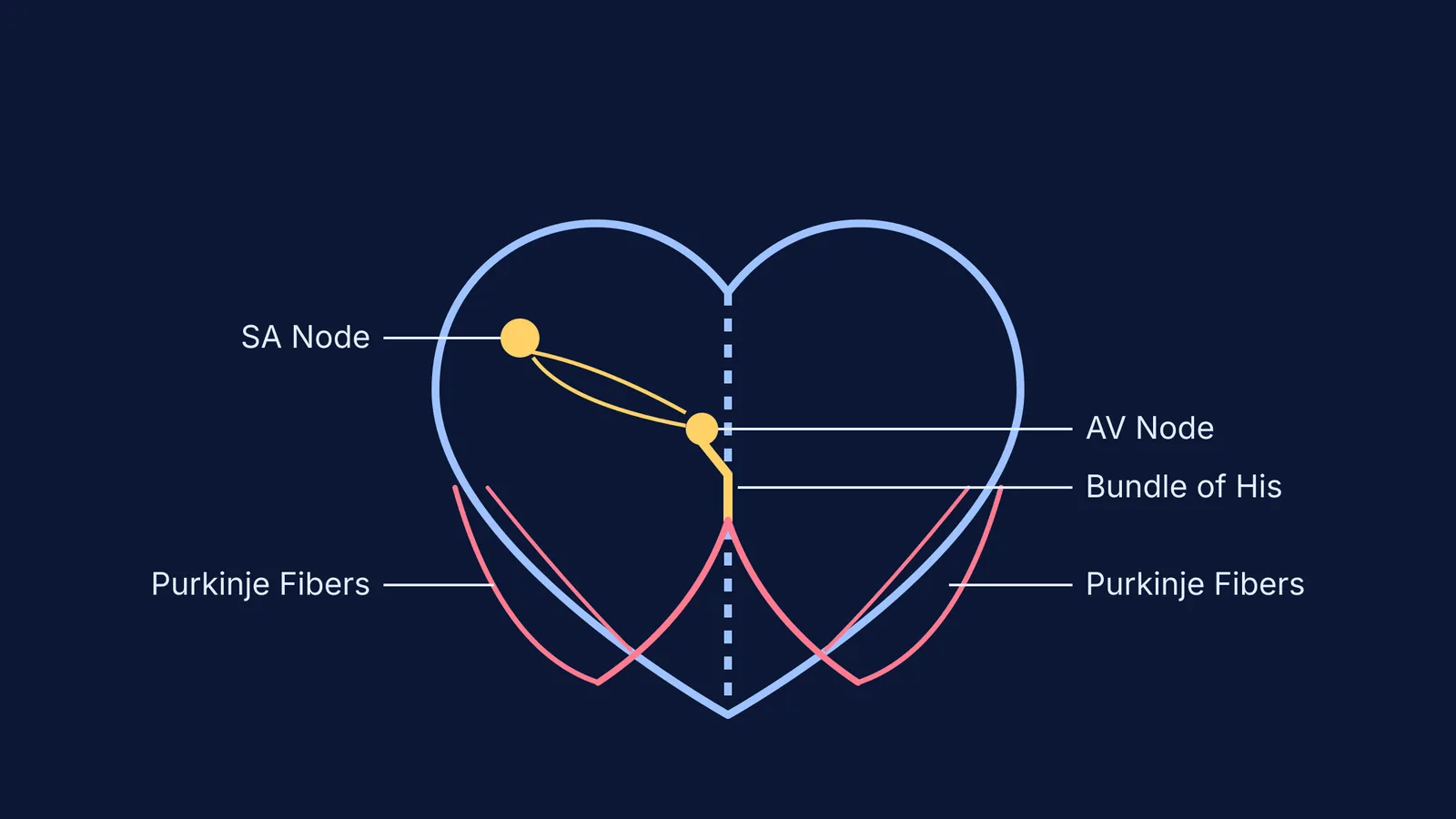
The heartbeat begins in the right atrium at the sinoatrial (SA) node. This crescent-shaped patch of specialized tissue acts as the heart's natural pacemaker. Unlike standard muscle cells, the cells of the SA node are auto-excitable; they do not have a stable resting phase and spontaneously generate electrical impulses.
The SA node generates action potentials at a rate of 70–75 per minute in a healthy, resting adult (NCERT). Because it fires faster than any other part of the conduction system, it dictates the overall heart rate. When the SA node fires, the electrical wave spreads rapidly across both the left and right atria, causing them to contract simultaneously and push blood downwards into the ventricles.
Why does the AV node delay the signal?
After sweeping across the atria, the electrical signal converges on the atrioventricular (AV) node, located at the lower left corner of the right atrium. Here, the conduction pathway does something unusual: it slows down.
The AV node delays the electrical impulse by approximately 0.09 seconds (Wikipedia). This brief pause is mechanically vital. It gives the atria enough time to finish their contraction and fully empty their blood into the ventricles (the "atrial kick") before the ventricles begin to contract. If the signal traveled without this delay, the upper and lower chambers would squeeze at the same time, severely reducing the volume of blood pumped out to the body.
How do the bundle of His and Purkinje fibers deliver the signal?
Once the delay is over, the signal must be delivered to the thick muscular walls of the ventricles as quickly as possible. From the AV node, the impulse enters the bundle of His (atrioventricular bundle), which is the only electrical bridge between the atria and the ventricles.
The bundle of His runs down the interventricular septum before splitting into the right and left bundle branches. These branches plunge toward the apex (the bottom tip) of the heart. Finally, they divide into a fine network of Purkinje fibers that spread upwards along the outer walls of the ventricles.
This specific routing—down the center and up the sides—ensures that the ventricular contraction begins at the bottom apex and wrings the blood upwards, directly into the aorta and pulmonary artery.
Tracing the conduction path for exams
When preparing for exams, keeping the sequence straight is essential. If you are memorising the pathway but struggling to picture the cardiac cycle, Fermi's AI tutor can help. Rather than just giving you the answer, it notices where you are stuck and runs the simulation of the heartbeat, letting you see exactly why the ventricles must squeeze from the bottom up.
You can also explore interactive models in the Fermi Concept Library to see the electrical wave in motion. To test your recall, use this four-step checklist:
SA node: Initiates the beat.
AV node: Pauses the signal.
Bundle of His: Carries the signal down the septum.
Purkinje fibers: Spreads the signal up the ventricular walls.
Understanding this pathway not only helps with biology questions but also connects to the history of medicine, such as the discovery made by Sunao Tawara and the atrioventricular node of the heart.


