

On this page
When you step out of bright sunlight into a dark cinema hall, your vision takes time to adjust. This everyday experience is driven by two distinct types of photoreceptor cells at the back of your eye: rods and cones. While both convert radiant energy into electrical signals for the brain, they are built for entirely different visual tasks.
What is the primary function of rods?
Rods are the specialists of scotopic vision—the ability to see in dim light. They are incredibly sensitive and can detect the smallest traces of light. This sensitivity comes from rhodopsin, a biological pigment that is exceptionally good at catching light but bleaches quickly when exposed to bright environments. When light hits rhodopsin, it changes shape, setting off a cascade of chemical reactions that acts like a biological amplifier.
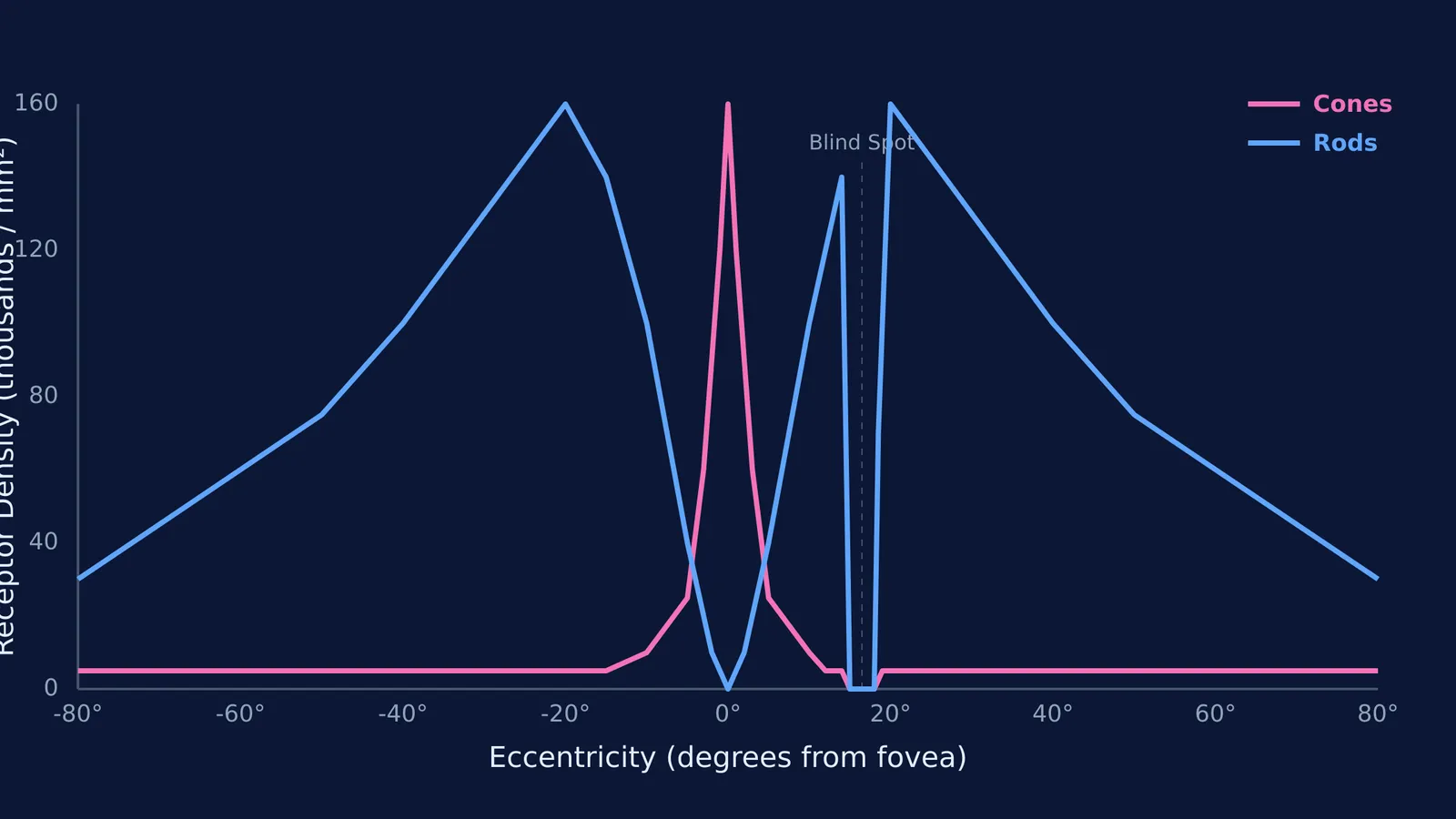
The human retina contains approximately 91 million rods (Purves et al., Neuroscience 2nd Ed.). Because they are mostly concentrated in the peripheral areas of the retina, away from the centre, rods are responsible for your peripheral vision. This acts as an early warning system, making rods excellent at detecting motion in the corner of your eye. To maximise their sensitivity, rods pool their signals. Many rods wire into a single nerve fibre, so a faint signal from any one rod is combined with others to ensure the brain registers it. However, this spatial pooling costs us clarity, which is why peripheral vision cannot resolve fine details.
How do cones process colour and detail?
Cones handle photopic vision—seeing in bright light. While they require significantly more light to activate than rods, they offer high-definition, colourful output. The retina contains around 4.5 million cones, and they are densely packed into a tiny central pit called the fovea (Purves et al., Neuroscience 2nd Ed.).
Unlike rods, cones contain three different types of photopsin pigments, each sensitive to a different wavelength of light: S-cones for short wavelengths (blue), M-cones for medium (green), and L-cones for long (red) (Review of Retinal Light Toxicity, PMC3144654). By comparing the overlapping signals from these three cone types, your brain constructs the full spectrum of colours you see. Colour blindness usually occurs when one of these cone types is absent or mutated.
In the fovea, the retina's inner cell layers are pulled aside so light hits the cones directly without scattering. Each cone here often has a direct, one-to-one connection with a nerve fibre (Purves et al., Neuroscience 2nd Ed.). This lack of pooling provides the maximum visual acuity required for threading a needle or reading a textbook; in fact, just 6 degrees away from the line of sight, visual acuity drops by 75% (Purves et al., Neuroscience 2nd Ed.). You can explore the entire structure of the eye in the Fermi Concept Library, which covers every concept in physics, chemistry, biology, mathematics and computing, each with its own lesson and its place in the map.
Why does it take time to see in the dark?
When you move from a brightly lit environment into a dark one, your cones stop working almost immediately because there are not enough photons to trigger them. Your rods, on the other hand, are temporarily useless because their rhodopsin stores have been completely "bleached" by the bright light.
It takes time for the retina to chemically rebuild the rhodopsin. This process, known as dark adaptation, takes significant time to complete fully. As the rhodopsin regenerates, your eyes become increasingly sensitive to the dim surroundings, eventually allowing you to navigate the dark room effectively.
Why do we only see grey shades at night?
As evening falls and light levels drop, the environment slowly loses its colour. Once the cones shut down, your vision shifts entirely to the rods.
Because rods contain only a single type of pigment, they can only measure the intensity of light, not its wavelength. They tell the brain how much light is present, but they cannot distinguish between red, blue, or green. As a result, your night vision is monochromatic, rendering the world in shades of grey.
Understanding how light interacts with biological pigments sits at the crossroads of physics and biology. If you are struggling to picture how wavelengths translate to colours, the Fermi Simulation Lab has interactive simulations for the ideas that are easier to see than to read. You can also revise the basics of the human eye and its defects or the laws of reflection, mirrors, and the human eye to see how lenses focus light onto the retina in the first place.
Rods and cones difference in retina functions: a quick summary
Sensitivity: Rods work in near-darkness; cones require bright light.
Colour vs B&W: Cones detect colour via three different opsins; rods only see shades of grey using rhodopsin.
Detail: Cones provide sharp, high-resolution images due to one-to-one nerve wiring; rods provide blurry, low-resolution shapes by pooling their signals.
Location: Cones are highly concentrated in the centre (the fovea); rods dominate the entire periphery.
Numbers: There are roughly 91 million rods and 4.5 million cones per eye.


