

On this page
In biology, Porifera (sponges) are famously simple: they have no organs, no true tissues, and no nervous system. Everything they do—eating, breathing, reproducing and removing waste—relies entirely on pumping a continuous stream of water through their bodies. This vital water-transport mechanism is called the canal system.
What is the path of water in Porifera?
The canal system drives water in a strict, one-way path through the sponge. Understanding this pathway is the foundation of sponge anatomy. Because sponges are sessile (they remain attached to the sea floor or rocks and cannot move to hunt or escape), they must bring the surrounding environment to them.
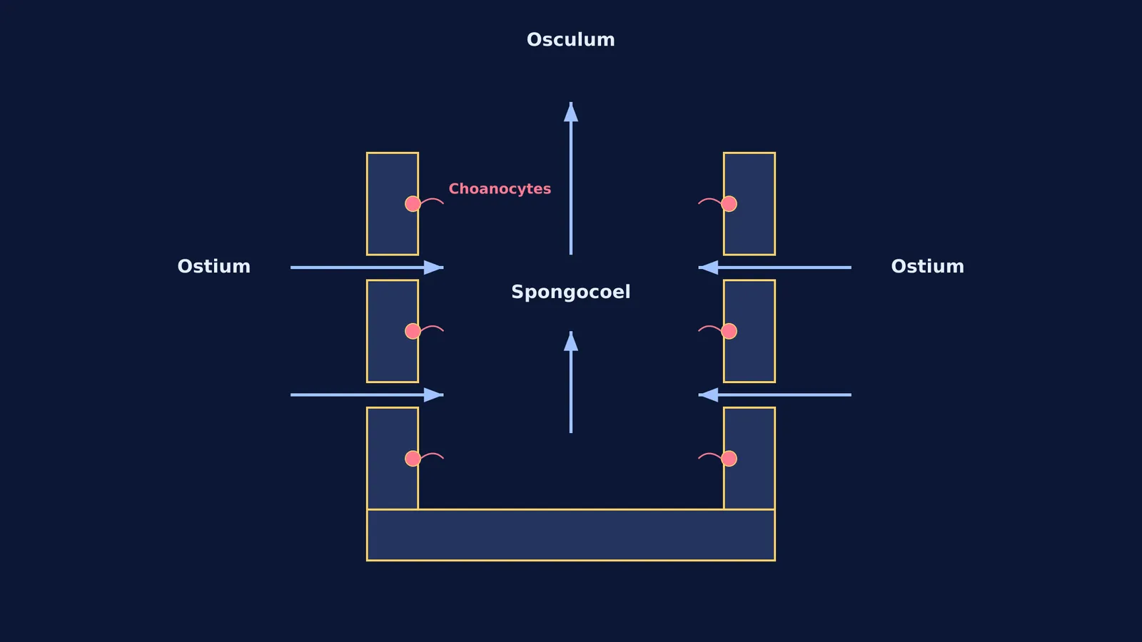
Ostia: Water from the surrounding ocean enters the sponge through thousands of minute pores called ostia, which are scattered all over the outer body wall.
Spongocoel: Once inside, the water collects in a central cavity known as the spongocoel.
Osculum: The water then travels upward and exits the sponge through a single, large opening at the top called the osculum.
This continuous current is the sponge's lifeline. The incoming water brings dissolved oxygen and microscopic food particles like plankton and bacteria. As the water passes through the spongocoel, the cells lining the cavity absorb oxygen and release carbon dioxide alongside metabolic wastes (such as ammonia), which are then safely flushed out into the ocean through the osculum.
What do choanocytes do?
Because sponges do not have a heart or muscles to move all this water, the current is driven entirely by specialised collar cells called choanocytes.
Choanocytes line the inner walls of the spongocoel and the various canals. Each choanocyte has a whip-like tail called a flagellum, surrounded by a collar of tiny, finger-like microvilli. (If you want to know more about how these structures propel movement, see our guide on cilia and flagella.)
The synchronized beating of these flagella creates a low-pressure zone that actively draws water in through the ostia and pushes it out through the osculum. As the water flows past the cell, the microvilli collar acts like a microscopic net, trapping suspended food particles. The choanocyte then engulfs these particles and digests them intracellularly.
Often, the choanocyte will hand off the partially digested food to amoebocytes—mobile, amoeba-like cells that wander through the jelly-like middle layer (the mesohyl) of the sponge. These amoebocytes complete the digestion process and deliver the resulting nutrients to the rest of the sponge's cells. They also gather waste products and dump them back into the outgoing water current.
The volume of water these tiny cells can move is substantial. Studies on marine demosponges show that a sponge can pump up to 35 millilitres of water per minute for every cubic centimetre of its tissue (Morganti et al., 2019). At this maximum rate, the sponge filters over 50,000 times its own volume in a single 24-hour period (Morganti et al., 2019).
How to study the canal system?
When revising Porifera for NEET-UG or Class 11 biology, it is easy to get caught up in the evolutionary progression of the canal systems. You will often see them divided into three main types:
Asconoid: The simplest type, found in small, tube-shaped sponges like *Leucosolenia*. Here, water flows directly through the ostia into a simple, undivided spongocoel.
Syconoid: As sponges grow larger, the simple asconoid design cannot provide enough surface area for choanocytes to pump sufficient water. In syconoid sponges, such as *Sycon*, the body wall is folded into radial canals. This folding dramatically increases the surface area lined by choanocytes without increasing the overall size of the spongocoel.
Leuconoid: The most complex and efficient design, found in the vast majority of sponges, including large bath sponges. The spongocoel is heavily reduced, replaced by an extensively branched network of canals that lead to small, round choanocyte chambers. This maximises the pumping and filtering capacity, allowing the sponge to grow to massive sizes.
Despite these structural differences, the fundamental physiological flow remains exactly the same in all three: water enters via the ostia, passes through a choanocyte-lined space (the spongocoel or a dedicated chamber), and exits via the osculum. Just as learning the defining features of viruses, viroids and lichens helps you tackle biological classification, remembering this simple path is the key to answering any exam question on Porifera.
If you struggle to picture how cells and tissues interact in 3D, tracing the path step-by-step is often the best way to memorise it. The Fermi Concept Library covers every concept in biology, each with its own lesson and its place in the map, helping you visualise the difference between a simple cavity and a complex folded canal. Once you understand the mechanism, you can test your recall with the Fermi Problem Library, which features worked problems with a step-by-step solver that explains the reasoning rather than printing an answer.


