

On this page
Every time you take a breath, your lungs expand and contract inside your rib cage. Without a specialised biological wrapper, the delicate tissue of the lungs would scrape against the hard bones and muscles of the chest wall with every breath, causing severe damage over time. To prevent this, the human body uses a double-layered barrier called the pleura to safely house the lungs.
What are the pleural membranes?
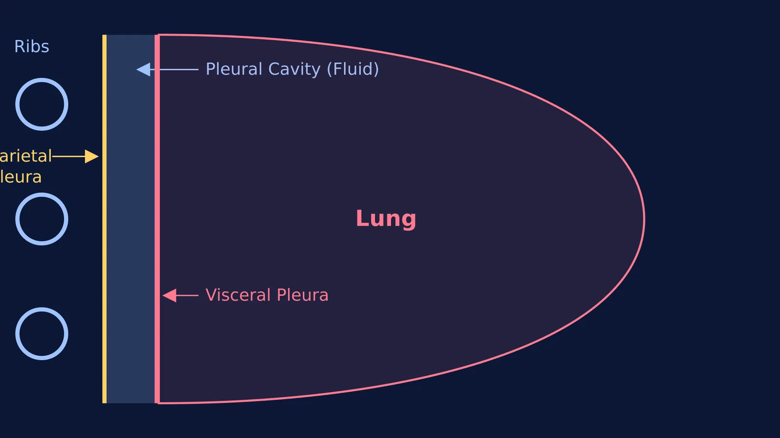
The lungs are enclosed in a continuous, folded covering made of a serous membrane. Think of it as pushing your fist into a partially inflated balloon: the side touching your hand is one layer, and the outside of the balloon is another. In the body, this structure forms two distinct layers:
Parietal pleura: This is the outer layer, which lines the inside of the thoracic cavity (the chest wall), the mediastinum, and the top of the diaphragm. According to a StatPearls clinical review, this layer is heavily vascularised and contains sensory nerve endings that can feel pain. When a respiratory issue causes sharp pain during a breath, it is typically the parietal pleura sending those signals.
Visceral pleura: This is the inner layer, which tightly adheres to the surface of the lungs, dipping deep into the fissures that separate the lobes. Unlike the outer membrane, it lacks sensory nerve fibres, meaning the lung surface itself does not register pain in the same way.
Between these two closely apposed layers lies a potential space known as the pleural cavity.
What is the function of pleural fluid?
The pleural cavity is not entirely empty; rather, it contains a small, meticulously maintained volume of liquid. Typically, this amounts to about 0.1 to 0.2 mL per kilogram of body weight in a healthy individual, as noted in clinical physiology outlines. This pleural fluid serves two vital mechanical functions for the respiratory system:
Lubrication: The fluid acts as a highly effective biological lubricant. It vastly reduces friction between the lung surface and the chest wall, allowing the parietal and visceral pleurae to glide past each other smoothly during the continuous cycles of inhalation and exhalation.
Surface tension and coupling: Just as a thin layer of water makes two glass slides stick together so tightly that they are hard to pull apart, the pleural fluid creates strong surface tension. This force couples the lungs directly to the chest wall. When your intercostal muscles and diaphragm contract to expand the thoracic cavity, the lungs are physically pulled outwards alongside them, ensuring efficient airflow.
In medical conditions such as pleurisy (inflammation of the pleura), this frictionless gliding is disrupted. The roughened membranes rub against each other during breathing, creating a characteristic "pleural friction rub" sound and causing sharp chest pain. Conversely, an overproduction or poor drainage of this liquid leads to a pleural effusion, where excess fluid compresses the lung and restricts breathing.
How is pleural fluid maintained?
The body ensures the pleural cavity never becomes too dry or too full. The fluid is continuously produced primarily by the capillaries of the parietal pleura. Once it has coated the membranes, the excess liquid is constantly reabsorbed into the lymphatic system through tiny openings called stomata, which are also located on the parietal side.
This continuous cycle of secretion and absorption guarantees that the exact volume needed for lubrication and surface tension is always present, keeping the pressure within the pleural space slightly negative compared to the atmosphere. This negative pressure is the mechanical secret that keeps your lungs from collapsing.
How do pleural membranes appear in exam questions?
In biology exams, questions on the human respiratory system frequently test your understanding of these membranes and the mechanics of breathing. To quickly and accurately answer these questions, keep this revision checklist in mind:
Location: The pleural fluid is strictly located between the parietal and visceral layers, never inside the lung tissue (alveoli) itself.
Roles: Always identify both primary functions in multiple-choice questions: reducing friction and coupling the lung to the chest wall via surface tension.
Origin and drainage: Recognise that the fluid is produced by the parietal pleura and drained by the lymphatic system.
Pressure: Remember that intrapleural pressure is always negative to prevent lung collapse.
If you are confused by how breathing mechanics and gas exchange relate, the Fermi Concept Library has every concept in physics, chemistry, biology, mathematics and computing, each with its own lesson and its place in the map. Reviewing how pulmonary circulation handles the oxygenated blood once it reaches the lungs, or studying the respiration pathway of the cells themselves, will give you a complete picture of the respiratory cycle from the nostrils to the mitochondria. For exam calculations involving gas exchange, knowing the respiratory quotient of different metabolic fuels is also an essential tool.


