

On this page
What are antenna pigments and light-harvesting complexes?
When sunlight strikes a leaf, reaction centres do not absorb every photon directly. Instead, chloroplasts surround each centre with an antenna network known as the light-harvesting complex (LHC). In the major Photosystem II complex (LHCII), whose crystal structure was resolved at 2.72 Å resolution by Liu et al. (2004), each protein monomer binds 14 chlorophylls (8 chlorophyll a and 6 chlorophyll b) alongside 4 carotenoids. Carotenoids absorb blue-green light between 400 nm and 500 nm while dissipating excess energy, and chlorophyll b captures wavelengths near 450 nm and 640 nm.
Antenna pigments do not transfer electrons or carry out redox chemistry. As reviewed by van Grondelle et al. (1994)90166-X), they transfer singlet excitation energy non-radiatively via Förster resonance energy transfer (FRET), a dipole-dipole mechanism operating at over 90% quantum efficiency. Excitation hops between pigments in 5 to 100 picoseconds, funnelling inward toward the thylakoid core. Decades earlier, Robert Hill's experiments with isolated chloroplasts showed that oxygen production occurs independently of carbon dioxide fixation, establishing that light capture is physically distinct from carbon reduction.
Why do P680 and P700 act as energy traps?
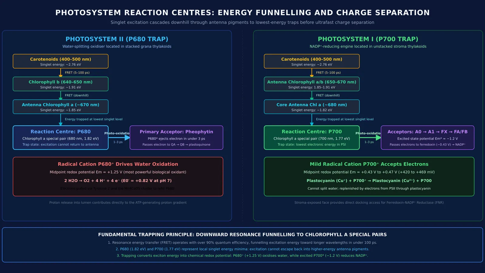
As explained in OpenStax Biology, light funnels according to thermodynamics: excitation energy flows downhill toward longer wavelengths and lower singlet states. Carotenoids absorb higher-energy photons near 450 nm (about 2.76 eV), transferring energy to chlorophyll b (around 650 nm, 1.91 eV), which feeds antenna chlorophyll a (around 670 nm, 1.85 eV). At the base of this cascade sits a specialised chlorophyll a pair called the reaction centre.
In Photosystem II, the reaction centre is P680, discovered by Horst Witt and colleagues in Döring et al. (1969) by tracking reversible flash-induced absorption changes at 680 nm. In Photosystem I, the reaction centre is P700, identified by Bessel Kok (1956)90172-x) through reversible photo-bleaching near 700 nm. Because P680 and P700 absorb at longer wavelengths than antenna pigments, their excited states occupy the lowest energy level in the complex. Once excitation arrives, it cannot hop backward. Within 1 to 3 picoseconds, this trapped excitation triggers charge separation, ejecting an electron to an acceptor.
For students mapping out photosynthetic pathways and bioenergetics, the Fermi Concept Library organizes every concept in physics, chemistry, biology, mathematics and computing, each with its own lesson and its place in the map.
What is the difference between P680 and P700?
While both reaction centres consist of chlorophyll a molecules, their distinct protein microenvironments tune them for different biochemical roles in the Z-scheme of photosynthesis in plants:
Oxidation potential and electron source: Upon ejecting an electron, P680 forms the radical cation P680⁺. Energetic measurements reviewed by Diner and Rappaport (2002) estimate the midpoint redox potential of P680/P680⁺ at approximately +1.25 V. This makes P680⁺ the strongest known biological oxidising agent, capable of extracting 4 electrons from 2 water molecules (+0.82 V at pH 7). Spectroelectrochemistry by Nakamura et al. (2005) shows P700/P700⁺ has a much milder redox potential between +0.42 V and +0.47 V, which cannot split water but readily accepts electrons from plastocyanin.
Reducing power and primary acceptors: Excited P680 transfers an electron to pheophytin, passing it down an electron transport chain to generate a proton gradient. Excited P700* reaches an extreme negative potential near −1.2 V, donating electrons through chlorophyll A0, phylloquinone A1, and iron-sulphur centres to ferredoxin (−0.43 V) to reduce NADP⁺ to NADPH.
Membrane location: Photosystem II complexes with P680 reside predominantly in stacked grana thylakoids, whereas Photosystem I with P700 sits in unstacked stroma thylakoids and grana margins where stroma enzymes like ferredoxin-NADP⁺ reductase are accessible.
How to master photosystems for NEET-UG?
Under the Senior Secondary Biology curriculum established by the Central Board of Secondary Education, plant bioenergetics questions frequently test the division of labor between antenna complexes, P680, and P700. Use this 4-step diagnostic checklist:
Verify energy flow: Confirm that excitation moves from shorter to longer wavelengths: carotenoids (~450 nm) to chlorophyll b (~650 nm) to chlorophyll a (~670 nm) and into the reaction centre (680 nm or 700 nm). Energy never flows backward under normal conditions.
Separate excitation from electron ejection: Antenna pigments pass singlet excitation energy non-radiatively without losing electrons. Charge separation occurs exclusively at the reaction centre chlorophyll a pair.
Trace the electron supply: P680⁺ extracts electrons from water via the oxygen-evolving complex, releasing molecular oxygen. In contrast, P700⁺ receives replacement electrons from Photosystem II via plastocyanin, or cycles them from ferredoxin during cyclic photophosphorylation.
Link light reactions to carbon fixation: Non-cyclic electron flow through P680 and P700 produces ATP and NADPH for the Calvin cycle. When downstream carbon reactions slow—such as when photorespiration wastes ATP and carbon—cells regulate photosystem activity to prevent oxidative damage.
When working through multi-step problems on photosynthetic electron transport, the Fermi Problem Library provides worked problems with a step-by-step solver that explains the reasoning rather than printing an answer.


