

On this page
Under a microscope, cross-sections of plant stems and roots can initially seem bewildering. Yet plant vascular anatomy follows two consistent architectural rules: how xylem and phloem are arranged relative to each other, and whether a dividing layer of meristematic cambium lies between them. Understanding this organization links the primary tissues formed by apical, intercalary and lateral meristems to each plant's capacity for secondary thickening.
What makes a vascular bundle open or closed?
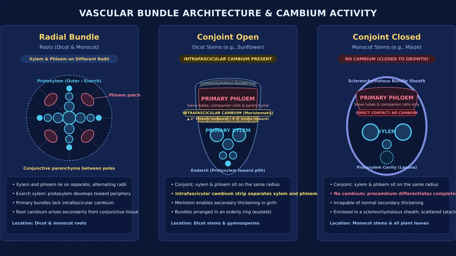
The defining test of an open versus a closed vascular bundle is the presence or absence of intrafascicular cambium. Primary vascular strands differentiate from procambium. In an open vascular bundle, a persistent strip of undifferentiated procambium remains between the primary xylem and primary phloem. As Ray F. Evert details in *Esau's Plant Anatomy* (DOI: 10.1002/0470047380), this residual meristem is called intrafascicular cambium, and it keeps the bundle open to secondary growth.
During secondary growth, this cambium divides to produce secondary xylem inward and secondary phloem outward. In *The Arabidopsis Book*, Kaisa Nieminen and colleagues (DOI: 10.1199/tab.0177) describe how this cylindrical meristem drives radial expansion in dicot shoots and hypocotyls. Furthermore, K. Fisher and S. Turner showed in *Current Biology* (DOI: 10.1016/j.cub.2007.05.049) that receptor kinase signaling maintains the spatial polarity of dividing vascular initials, keeping xylem and phloem organized.
In a closed vascular bundle, every procambial cell differentiates completely into primary conducting tissues. Without cambium, the bundle cannot produce secondary tissues. Closed bundles characterize monocot stems and leaves, where bundles are typically enclosed by a sclerenchymatous sheath.
If you need to connect tissue differentiation to developmental stages, the Fermi Concept Library covers every concept in biology, each with its own lesson and its place in the map.
How do radial and conjoint vascular bundles differ?
Radial versus conjoint arrangements describe whether xylem and phloem share a single radius or occupy different radii.
Radial bundles: In roots, xylem and phloem lie on separate, alternating radii, separated by parenchymatous conjunctive tissue. Roots exhibit exarch xylem development, where protoxylem lies toward the periphery and metaxylem develops toward the centre.
Conjoint bundles: In stems and leaves, xylem and phloem sit together along the same radius. Stems show endarch xylem development: protoxylem faces the central pith, while metaxylem faces the cortex.
Conjoint bundles include two main forms:
Collateral: Phloem lies on only one side (the outer abaxial margin) of the xylem. Dicot stems have collateral open bundles; monocot stems have collateral closed bundles.
Bicollateral: Phloem occurs on both outer and inner margins of the xylem, flanked by two cambium strips. Cankui Zhang and colleagues noted in *Plant Physiology* (DOI: 10.1104/pp.112.194431) that this open bicollateral layout provides dual phloem pathways in Cucurbitaceae stems, such as pumpkin.
Even when plants form underground and subaerial stem modifications like rhizomes or tendrils, their internal tissues retain these conjoint vascular arrangements.
Where does each vascular bundle type occur in plants?
Recognizing where each bundle type occurs clarifies cross-sections across plant organs:
Dicot stems: Conjoint, collateral (or bicollateral), open bundles arranged in an orderly ring (eustele) around a central pith. Medullary ray parenchyma dedifferentiates into interfascicular cambium, completing the cambial ring.
Monocot stems: Conjoint, collateral, closed bundles scattered through ground parenchyma (atactostele). Outer bundles are smaller and densely crowded; inner bundles are larger and widely spaced.
Roots: Radial bundles in both dicots and monocots. As documented in *Esau's Plant Anatomy* (DOI: 10.1002/0470047380), dicot roots typically have 2 to 4 xylem arches (diarch to tetrarch), whereas monocot roots are polyarch with more than 6 arches.
Leaves: Conjoint, collateral, closed bundles in both dicots and monocots.
How can you identify vascular bundle types in exam problems?
When examining cross-sections in exam questions, use this three-step identification process:
Check radius alignment: If xylem and phloem alternate along different radii, the organ is a root with radial bundles. If they share the same radius, the organ is a shoot with conjoint bundles.
Check for cambium: A tidy band of rectangular meristematic cells between xylem and phloem marks an open bundle. Direct contact between xylem and phloem without meristem marks a closed bundle.
Check section distribution: A single organized ring around a distinct pith indicates a dicot stem. Scattered bundles embedded throughout ground tissue indicate a monocot stem.
When practicing plant anatomy questions, the Fermi Problem Library has worked problems with a step-by-step solver that explains the reasoning rather than printing an answer. If an unfamiliar cross-section appears confusing, Fermi's AI tutor works out what you already know, builds a plan for the session, and checks understanding as it goes.


