

On this page
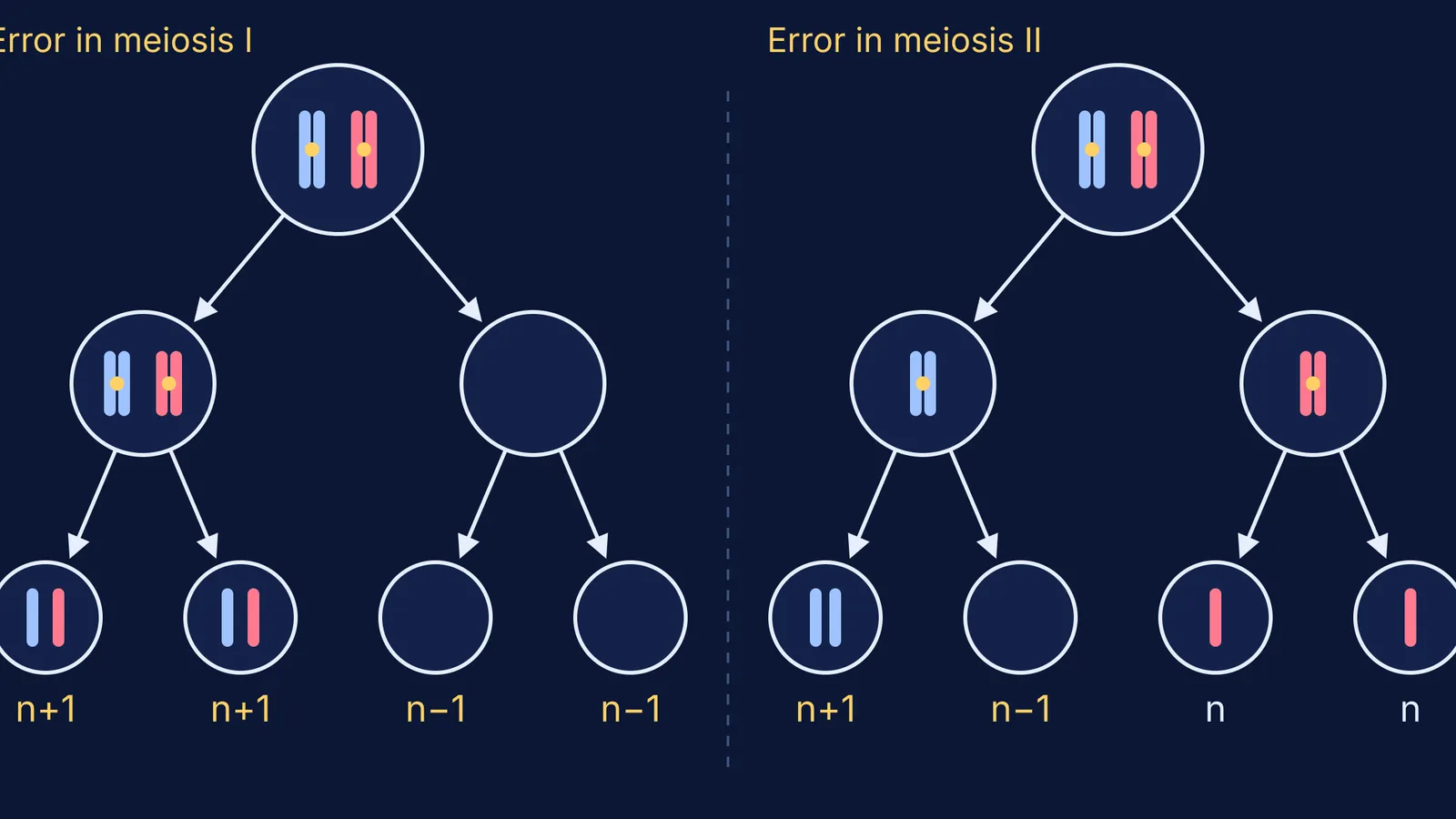
Your question shows a parent cell with one pair of homologous chromosomes and asks how many of the four gametes are abnormal. The answer depends on which division went wrong.
What is nondisjunction?
Nondisjunction is an error in cell division that leaves a reproductive cell with an abnormal number of chromosomes, as MedlinePlus Genetics defines it. In meiosis I the things that should separate are homologous chromosomes. In meiosis II they are sister chromatids. For a refresher, see our notes on stages and chromosome counts in mitosis and meiosis.
Aneuploidy (trisomy or monosomy) occurs in at least 5% of all clinically recognised pregnancies, according to a review by Terry Hassold and Patricia Hunt in Nature Reviews Genetics. The same review calls it the leading genetic cause of pregnancy loss.
What gametes form after nondisjunction in meiosis I?
If homologous chromosomes fail to separate during meiosis I, the result is two gametes with two copies of that chromosome and two gametes that lack it (LibreTexts Biology).
Anaphase I: both homologs move to the same pole.
One cell has the whole pair, the other has neither.
Meiosis II runs normally in both.
Result: n+1, n+1, n−1, n−1. All four gametes are abnormal.
Each n+1 gamete carries two different homologs, one that came from the parent's mother and one from the parent's father.
What gametes form after nondisjunction in meiosis II?
If sister chromatids fail to separate during meiosis II, the result is one gamete that lacks the chromosome, two normal gametes and one gamete with two copies (LibreTexts Biology).
Meiosis I is normal: each cell gets one replicated homolog.
In one of the two cells, the sister chromatids stay together at anaphase II.
The other cell divides normally.
Result: n+1, n−1, n, n. Half the gametes are normal.
Here the extra copy is a sister chromatid, so the n+1 gamete carries two copies of the same homolog.
How do n+1 and n−1 gametes lead to trisomy and monosomy?
At fertilisation the faulty gamete meets a normal one (n). An n+1 gamete gives a 2n+1 zygote, a trisomy. An n−1 gamete gives a 2n−1 zygote, a monosomy.
Trisomy 21 (Down syndrome): an extra chromosome 21. Down syndrome occurs in about 1 in 700 newborns, according to MedlinePlus Genetics.
47,XXY (Klinefelter syndrome): an egg or sperm carries an extra X. It affects about 1 in 650 male newborns (MedlinePlus Genetics).
45,X (monosomy X): an egg or sperm lacks a sex chromosome. About half of people with Turner syndrome have monosomy X; Turner syndrome occurs in about 1 in 2,000 female newborns (MedlinePlus Genetics).
Why are the monosomy examples nearly always about X? Human zygotes missing one copy of any autosome fail to develop to birth, as the LibreTexts chapter on chromosomal disorders explains.
Which parent, and which division? A 2007 review by Hassold, Hall and Hunt in Human Molecular Genetics states that most aneuploidy comes from errors in maternal meiosis I, and that maternal age is a risk factor for most, if not all, human trisomies. For how these appear in pedigree and exam questions, see our guide to sex determination, pedigrees and chromosomal disorders.
How can you tell if the error was in meiosis I or II?
A meiosis I error passes on two different homologs; a meiosis II error passes on two copies of the same one. A marker gene shows which. Exam questions usually ignore crossing over, which can blur this (see linkage and crossing over).
Worked example. Write C for the normal-vision allele and c for red-green colour blindness. The mother is a carrier (XC Xc) and the father has normal vision (XC Y). They have a colour-blind son who is XXY.
A colour-blind XXY son must be Xc Xc Y.
The Y came from the father, so both X chromosomes came from the mother.
Both carry c, but the mother has only one Xc. Two identical copies means sister chromatids stayed together.
Answer: nondisjunction in the mother, in meiosis II.
Now change one fact: the XXY son has normal vision and is XC Xc Y. That could be the mother in meiosis I (both of her different X chromosomes), or the father in meiosis I (an XC Y sperm) meeting the mother's Xc. A full answer names both.
If the method trips you up, the Fermi Problem Library has worked problems with a step-by-step solver that explains the reasoning rather than printing an answer. To revisit homologs, chromatids and centromeres one at a time, the Fermi Concept Library gives each concept its own lesson and its place in the map.
This post explains exam biology. If your family is dealing with a real test result, a doctor or genetic counsellor is the right person to ask.


