On this page
The kidneys continuously filter blood to produce urine, but the human body does not constantly leak. Instead, urine is stored in the bladder and released only when appropriate. This entire process is controlled by the micturition reflex. Understanding the neurophysiology of micturition is a key part of human anatomy. It is not merely a simple loop in the spinal cord; it involves an intricate balance between the autonomic nervous system and the brainstem. While the kidneys determine urine concentration—a process you can explore in our guide to regulating kidney function: ADH, the RAAS and ANF explained—the bladder serves as a highly compliant holding tank, expanding smoothly until it receives the signal to empty.
How does the bladder store urine?
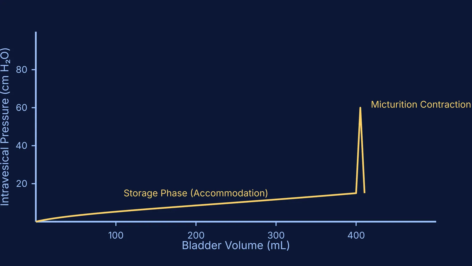
The walls of the urinary bladder are lined with a specialised smooth muscle known as the detrusor. During the storage phase, the detrusor muscle relaxes to allow the bladder to expand as it fills with urine. This property is known as accommodation, meaning the bladder volume increases while the internal pressure remains remarkably low.
This phase is controlled primarily by the sympathetic nervous system. Sympathetic nerve fibres originating from the T11 to L2 segments of the spinal cord release noradrenaline (Fowler et al., 2008). This neurotransmitter serves a dual purpose: it acts on β3-adrenergic receptors to keep the detrusor muscle relaxed, and it stimulates α1-adrenergic receptors in the bladder neck and internal urethral sphincter to keep them tightly closed. This guarantees that as long as the bladder is storing urine, the exit route is physically blocked.
How do stretch receptors signal the urge to void?
As the adult bladder fills to roughly 150 to 250 mL, you will usually feel the first sensation of filling (de Groat, Griffiths & Yoshimura, 2015). This sensation is picked up by stretch receptors—specifically, myelinated Aδ-fibres located within the detrusor muscle wall. These sensory nerves detect the physical distension of the bladder tissue.
As the volume continues to increase toward 300 to 500 mL, these stretch receptors fire more rapidly. Their signals travel along the pelvic nerve into the sacral spinal cord, and then ascend to relay stations in the brain, including the Periaqueductal Grey (PAG). The PAG acts as a sensory hub, forwarding the information to higher cortical centres so that you consciously register a strong urge to void. This waste removal process is standard for mammals, which you can contrast with other species in our post on ammonotelic, ureotelic and uricotelic animals: why waste differs.
What triggers the micturition reflex?
The micturition reflex is the coordinated neurological switch from storing urine to expelling it. The master control for this switch is located in the brainstem and is called the Pontine Micturition Center (PMC).
When the stretch receptors signal that the bladder is full, and the brain decides it is an appropriate time and place to urinate, the PMC sends descending signals to initiate voiding (Fowler et al., 2008). It flips the switch by inhibiting the sympathetic storage pathways and activating the parasympathetic nervous system. Parasympathetic nerve fibres from the S2 to S4 segments of the spinal cord release acetylcholine, causing the detrusor muscle to forcefully contract. At the same time, the internal urethral sphincter relaxes, allowing urine to flow out into the urethra. If you struggle to keep these competing autonomic functions straight, you can find a clear lesson on them in the Fermi Concept Library.
Why can you hold your urine?
The reflex to empty the bladder is powerful, but healthy adults can override it. This is possible because the external urethral sphincter, unlike the internal one, is composed of skeletal muscle. Skeletal muscle is under voluntary, somatic control.
This voluntary control is managed by the pudendal nerve, which also originates from the S2 to S4 segments of the spinal cord (Fowler et al., 2008). If you need to delay urination, your brain's prefrontal cortex sends an inhibitory signal to the PMC and a stimulatory signal down the pudendal nerve, commanding the external sphincter to remain firmly clamped shut. This voluntary "guarding reflex" gives you the time needed to reach a bathroom. To better understand how skeletal and smooth muscles differ, you can run interactive simulations in the Fermi Simulation Lab.